Introduction
Temporomandibular dysfunction (TMD) is a term that describes a number of clinical problems that involve the hard and soft tissue structures of the temporomandibular joint (TMJ) (1). Variety symptoms are related to TMD including clicking or grating within the joint, mechanical restrictions, headache, neck pain, or stiffness (2). In the general population, more than 25% have developed TMD and nearly two-thirds of the TMD patients are treated by dentists or physicians (3). However, analyzing TMJs’ condition and evaluating the treatment progress with TMD patients is very challenging.
Most TMD patient has clinical issues with mechanical restrictions such as limited jaw opening capacity and deviations in the movement patterns of the mandible (4). The study of jaw motion is essential to the management of TMDs. The need to duplicate the mandibular movements extra-orally led to the employment of various methods to record and analyze them (5, 6). There are several limitations in using a physical articulator with the mounted cast to evaluate the TMJs’ condition as articulators are unable to record real-life dynamics of occlusion during mandibular movements. Also, the accuracy of transforming the mandibular action is questionable (7).
Currently, a new mandibular tracing system can be used to evaluate the TMJs’ condition and associate the TMD problem with jaw movement. SICAT JMT + (Sirona, Bensheim, Germany) is the jaw motion tracker used to record and measure jaw position and movement in conjunction with cone beam computed tomography (CBCT) and digital intraoral impression (8). The system converts the propagation times of multiple acoustic signals into spatial information using optoelectronic systems. The para-occlusal attachment of the ultrasonic transmitter is to be attached to the patient and blocks occlusal bite relationships. Then, the jaw movement is optically tracked by sensors in the digital face bow (9). The CBCT data are also used to image the TMJs, which shows the observation of boney joint structures in all three planes in addition to the possible image manipulation at different depths and three−dimensional reconstructions (10). Meanwhile, the digital impression data can be used to evaluate the occlusal condition of TMD patients.
At present, there are few reports of the conjunction with digital mandibular tracing and TMD patients. The purpose of this case report was to present a digital tracing technique used to evaluate a patient with temporomandibular joint dysfunction.
Report of case
Chief complaints
A 65-year-old Caucasian female presented to the prosthodontic postdoctoral clinic of Boston University Henry M. Goldman School of Dental Medicine. Her chief complaint was feeling like her bite is off after having crowns on her lower front teeth and chronic jaw pain in the TMJ area. She also complained about headaches and neck and shoulder pain for a duration of 12 months.
Medical and dental history
The patient had a history of orthodontic treatment at age 15 and stopped treatment one year later. The patient described the treatment for moving her bite from Angle Occlusal Class III to Class I. At age 30, tooth #30 (mandibular first molar, ADA system) was extracted without any restorations. The patient reported a head-on collision at age 40 in which she hit her face on a basketball and lost tooth #25 without mentioning any fracture or trauma at the bone. A six-unit fix partial denture from #27–26 × 24–23–22 was fabricated. The patient reported the first jaw pain after this treatment, and the pain subsided after a few months. At age 50, the patient was treated with veneers from #5–11 because of cosmetic issues. Three years later, the veneered teeth #8, 9, and 10 were broken, and porcelain crowns were fabricated. Three years later, teeth #9 and #10 porcelain crowns were broken, and root canal treatment, cast post and core, and porcelain fused to metal crowns were fabricated. The patient complained of jaw pain after every dental treatment, and the pain subsided after a few months.
The patient reported no current prescription and no over-the-counter use of medication prior to her visit. The patient was diagnosed with TMD by a physician who also gave a preliminary diagnosis of fibromyalgia and therefore referred her to a dentist.
Intra-oral and extra-oral exam (Figures 1, 2).
During the exam, the patient exhibited a symmetric maxillofacial structure in the frontal view and an average lower facial height. Extra-orally, her profile was slightly convex with an extrusive lower lip. The intra-oral exam found tooth #3 super-erupted, multiple broken restorations, facial gingival recession, and number of teeth rotations. Her upper dental midline was coincident with the face midline. The lower dental midline deviated 2 mm to the left.
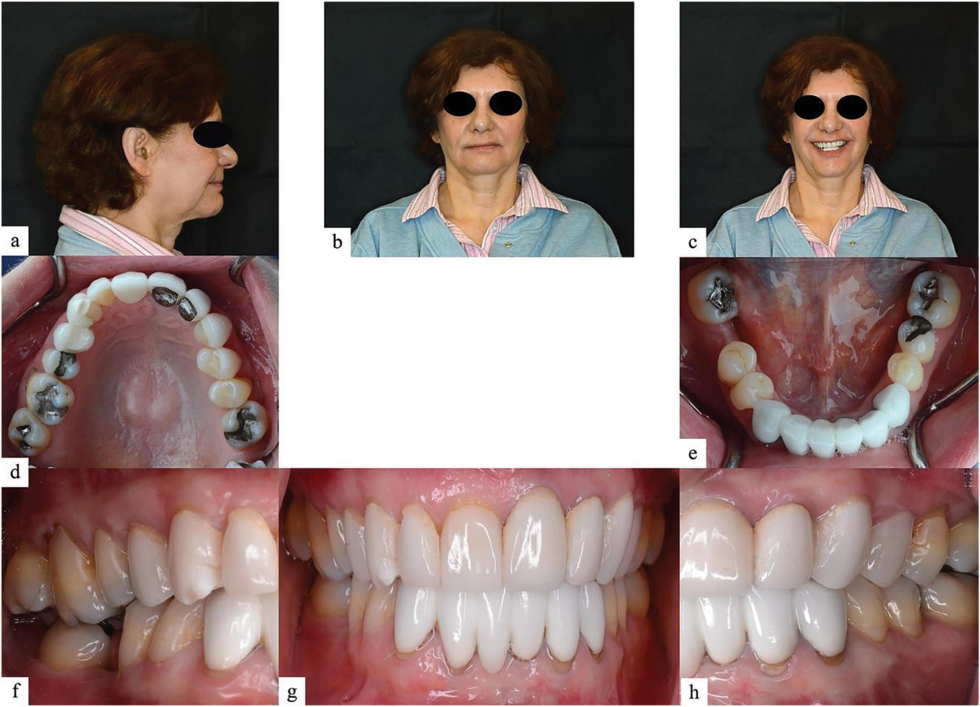
Figure 1. Facial and intraoral photographs of the patient. (a) Facial profile, (b) facial front view, (c) facial front smiling, (d) maxillary arch occlusal view, (e) mandibular arch occlusal view, (f) intraoral right lateral view in maximum intercuspation, (g) intraoral frontal view in maximum intercuspation, and (h) intraoral left lateral view in maximum intercuspation.
Figure 2. Intraoral photographs of jaw movements. (a) Right lateral view in right laterotrusion, (b) frontal view in right laterotrusion, (c) left lateral view in left mediotrusion, (d) right lateral view in right mediotrusion, (e) frontal view in left laterotrusion, (f) left lateral view in left laterotrusion, (g) right lateral view in protrusion, (h) frontal view in protrusion, and (i) left lateral view in protrusion.
The occlusal plane on the right side was uneven with the super-erupted tooth #3. The occlusal plane on the left side was flat. The patient displayed Angle Class I molar and canine relationship. Group-function articulation transitioning to canine-protected articulation was presented on both sides. On the right side, non-working contacts were presented between teeth #3 and 31 while in the left lateral excursion. In protrusion, there was no occlusion of the posterior teeth with #8 and 25, and 26 in contact. In general, the patient had 2 mm overbite, 1mm overjet, and 2 mm interocclusal rest space.
The patient mentioned that she had been suffering from TMJ issues her whole life, including symptoms of clicking and pain. History revealed that her jaw pain was on the left side with clicking and popping with jaw range of motion. The patient reported constant pain in the left frontal aspect of her head that she rated at a 7/10 on a numeric pain scale. She had a pain increase in chewing, and nothing reduced it. The patient also complained about a migraine-type headache, neck pain with tiredness, sore neck muscles, and shoulder aches.
Cone beam computed tomography and mandibular motion tracking (Figure 3).
Cone beam computed tomography was taken using Galileos Comfort (Sirona, Galileos, Bensheim, Germany) with a patient bite on the Fusion Bite tray, which had eight radiopaque markers as landmarks for the superimposition of CBCT and JMT data. SICAT JMT+ was used to record patient mandibular movement strictly following manufacturer manuals (11). The patient’s jaw movement was recorded including jaw opening and closing movements to and from the habitual intercuspal position, lateral and protrusive movements, border movement, and chewing.

Figure 3. SICAT JMT+ components. (a) Lower jaw sensor and facebow type 13 R, (b) T-attachment, and (c) Fusion Bite Tray.
The CBCT and JMT data were loaded into the SICAT Function software. Mandibular and glenoid fossa segmentation was performed, and the software extracted CBCT data and merged it with the corresponding jaw motion data. The system then presents a 3D image of the patient−specific mandibular movement on the screen.
Full−arch optical impressions of the patient were obtained with an intraoral scanner (Sirona, CEREC Omnicam). The images were imported and merged with the CBCT data simultaneously with the mandibular and condylar segmentation in SICAT Function software. This procedure gives the possibility to assess the dynamic occlusion and mandibular movement in interaction with TMJ function.
Digital data analyzing
First, CBCT images in the TMJ area were used to analyze the shape and potential wear in the condyle and fossa. The patient’s condyles were not in a bilaterally symmetric shape. The right condyle was slightly flat compared with the left on the anterior surface, and the fossa was bilaterally symmetric, which indicated wear and tear of the condyles (Figure 4).

Figure 4. Temporomandibular joint (TMJ) images from cone beam computed tomography (CBCT). (a) Axial view from above, (b) right cross-sectional view, (c) left cross-sectional view, (d) right tangential view, and (e) left tangential view.
Next, the jaw opening-closing movements and lateral-protrusive movements were analyzed. The results showed the maximal mouth opening at 38.9 mm, slightly smaller than the average range (12). The lateral right movement was 10.8 mm, and the lateral left movement was 8.4 mm in incisal. This result supported the clinical findings of the non-working contact on the right side. The protrusion movement was 6.8 mm, which was significantly smaller than the normal range and indicated that the patient had limited protrusion movement (Figure 5).

Figure 5. Parameters of jaw movement.
Finally, the border movement data were used to analyze the vibration and stabilization of the jaw. In a segmental view, this patient showed asymmetry in border movement. In the right border movement, the patient could not move smoothly and evenly. This result marched the asymmetric shape of the condyle and revealed the damage of the TMJs in both bone and connecting tissue. Meanwhile, the patient could not repeat the jaw motion properly, which was another evidence leading to the dysfunction of TMJs (Figure 6).

Figure 6. Front view of recorded border movement.
Discussion
This case study is probably one of the first in the literature to describe collaborative approaches to evaluate TMD with mandibular movement tracing. The patient in this study responded well to the evaluation and diagnosed visits without any additional complaints.
With the help of mandibular tracing data, we were able to find the cause of this patient’s TMD problem so a proper treatment plan could be presented to the patient. The tracing data presented evidence for establishing a treatment plan and also fulfilling the qualification of evident-base treatment. According to the patent’s dental history, plus clinical examination, and mandibular motion evaluation, the cause of this patient’s TMD problem is as follows:
1. Unfinished orthodontic treatment at an early age changes the growth pattern of the mandibular jaw. As this therapy modified the patient‘s occlusal anatomy from Class III to Class I in the mandibular growth period, the maxillary locked proper mandibular matrix, and intra-matrix rotation in growth (13). Limited protrusion movement is substantial evidence that shows in tracing results. Overgrowth of the chin and long mandibular anterior teeth are also evident. Although the patient did not mention any complaint about this treatment, it played an important role as the original reason for the TMD problems.
2. Improper mandibular anterior restorations are the trigger of jaw pain. The abnormal height of the mandibular anterior restoration limits the mandibular movements, which causes the patient’s first jaw pain complaint. The broken restorations in the maxillary anterior teeth also suggest this problem. As a result, TMD complaints started after the delivery of mandibular anterior fix partial denture.
3. Molar super-eruption and rotation causes improper contact on the non-working side. Unsupported teeth #3 and #31 created abnormal contacts in the non-working side, which forces the mandibular jaw to hit and slide when closing. In tracing data, it clearly shows the uncertain movement of the patient’s closing motion, which is strong evidence for this problem. Also, the unbalanced lateral movement range indicates that these improper contacts enhance the damage of the TMJs.
Depending on the clinical findings and analysis of the problem of TMD, the following treatment options can be considered:
1. An occlusal appliance should be considered to reposition the mandibular from the lock of the maxillary. Anterior repositioning appliance (ARA) without anterior teeth contact is one of the recommended appliances. ARA is used primarily for the management of TMD and was first described by Farrar in the 1970 s (14). It has been reported that ARA can relieve specific symptoms of pain in TMD (15). The aim of using ARA in this patient is to gain support for posterior teeth and release the mandibular jaw from the lock position. It can reestablish normal disc-condyle-fossa relationships through the use of therapeutic mandibular positions that are forward to maximum intercuspation.
2. Occlusal adjustment for contact equilibration contact is another treatment option. Removing the abnormal contact in occlusal and jaw movement can stop the hit and slight effect for continuous damage to the TMJs. With the help of jaw motion tracking, real-time monitoring is possible when operating this treatment. Also, tracing can be taken before and after treatment to evaluate the treatment result.
3. Replacing the improper restoration and establishing a stable occlusion are also options for this patient’s treatment.
Limitations
One of the limitations of this case report is that we did not evaluate the patient jaw and connect tissue condition with magnetic resonance imaging, and we did not record the patient’s muscle reaction such as electromyography. There are also several diagnostic and analyzing methods that can be used to evaluate TMD patients. This makes it difficult to determine what was the effective evaluation method and if it was necessary to perform a combination of all methods. Further treatment options and post-treatment evaluation should be present in this case, and more research and case studies should be generated with a specific category of TMD, like an articular disk displacement, and fewer manual therapies with the dental splint to isolate a better method.
Conclusion
This case report described how a mandibular tracing system could be used in evaluating temporomandibular joint dysfunction cases. Digital mandibular tracing systems can help the dentist to diagnose and analyze the causes of TMD specifically and individually for these patients. Association between digital mandibular tracing and TMD patient radiographic images may also help to provide a quick resolution and management for patients with TMD.
References
1. Cuccia A, Caradonna C, Caradonna D. Manual therapy of the mandibular accessory ligaments for the management of temporomandibular joint disorders. J Am Osteopath Assoc. (2011) 111:102–12.
3. McNeely M, Olivo S, Magee D. A systematic review of the effectiveness of physical therapy interventions for temporomandibular disorders. Phys Ther. (2006) 86:710–25.
4. Riley J, Gilbert G. Orofacial pain symptoms: an interaction between age and sex. Pain. (2001) 90:245–56.
5. Slavicek R. Relationship between occlusion and temporomandibular disorders: implications for the gnathologist. Am J Orthod Dentofacial Orthop. (2011) 139:10, 12, 14.
6. Costa H, Slavicek R, Sato S. A computerized tomography study of the morphological interrelationship between the temporal bones and the craniofacial complex. J Anat. (2012) 220:544–54.
7. Gsellmann B, Schmid-Schwap M, Piehslinger E, Slavicek R. Lengths of condylar pathways measured with computerized axiography (CADIAX) and occlusal index in patients and volunteers. J Oral Rehabil. (1998) 25:146–52.
8. Aslanidou K, Kau C, Vlachos C, Saleh T. The fabrication of a customized occlusal splint based on the merging of dynamic jaw tracking records, cone beam computed tomography, and CAD-CAM digital impression. J Orthod Sci. (2017) 6:104–9.
9. Hanssen N, Ruge S, Kordass B. SICAT function: Anatomical real-dynamic articulation by merging cone beam computed tomography and jaw motion tracking data. Int J Comput Dent. (2014) 17: 65–74.
10. Ferreira L, Grossmann E, Januzzi E, de Paula M, Carvalho A. Diagnosis of temporomandibular joint disorders: Indication of imaging exams. Braz J Otorhinolaryngol. (2016) 82:341–52.
11. He S, Kau C, Liao L, Kinderknecht K, Ow A, Saleh T. The use of a dynamic real-time jaw tracking device and cone beam computed tomography simulation. Ann Maxillofac Surg. (2016) 6: 113–9.
12. Faulkner K, Atkinson H. Mandibular movement in lateral excursions. J Oral Rehabil. (1984) 11:103–9.
13. Björk A, Skieller V. Normal and abnormal growth of the mandible. A synthesis of longitudinal cephalometric implant studies over a period of 25 years. Eur J Orthod. (1983) 5:1–46.
14. Farrar W. Diagnosis and treatment of anterior dislocation of the articular disc. N Y J Dent. (1971) 41:348–51.