Introduction
Globe subluxation (GSL) is a medical emergency that can occur suddenly, voluntarily, or by accident. Among them, spontaneous GSL is rare, especially in children, and it’s an anxious situation for both the patient and the ophthalmologist. GSL was first reported by Tucker in 1907. (1) Different literature searches in Google Scholar, Scopus, Medline, and PubMed showed 36 cases of spontaneous GSL to date (2–11). According to the review, the affected patients range in age from 8 months to 73 years (2).
In children, GSL mostly occurs due to trauma, and spontaneous GSL is very rare (10). Here we report a case of spontaneous GSL in a 14-day-old baby who had bilateral proptosis since birth, a very small anterior fontanel, no limb anomalies, and a shallow orbit in a computerized tomography (CT) scan, which are the characteristic features of craniosynostosis, especially in favor of Cruzuon syndrome. This was probably the first reported neonatal case of spontaneous GSL.
Case report
A 14-day-old-male baby was presented with his parent with the complaints of sudden painful outward protrusion of right eye which became red and swollen for 10 days. Parents also noticed that the baby’s eyes were slightly proptosed since birth. The boy was delivered by normal vaginal delivery with a birth weight of 2,900 g. He was the second child of his parents, and there was no family history of any facial anomalies.
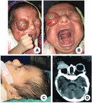
On examination, the baby was toxic, and gross facial asymmetry was present. The right eye was outside the orbital rim with conjunctival congestion and chemosis, and the baby was unable to close the eye. The cornea of the right eye was severely dry with surrounding infiltration (Figure 1A). There was no visual potential in the right eye. His left eye was slightly proptosed as well.

Figure 1. Clinical presentation. (A) Right globe subluxation with corneal exposure keratopathy and conjunctival swelling. (B) High arched palate with very upper lip. (C) Frontal bossing, depressed nasal root with curved tip of nose and left eye proptosis. (D) CT scan showing no mass lesion in right orbit with bilateral shallow orbit.
The baby had facial abnormalities like frontal bossing, hypertelorism, a flat nasal root with a mildly curved nose tip, a highly arched palate, and a very thin upper lip (Figures 1B, C). His head circumference was 36 cm, and the anterior fontanel was 0.5 cm, which was smaller than normal size with no abnormalities in the extremities.
His complete blood count and thyroid profile were normal. A CT scan of the orbit and brain showed no mass lesion in the right orbit, and the right globe was totally out of orbit. Both orbits were shallow, and the left eye was proptosed radiologically (Figure 1D). His left eye and brain were normal except for hypomyelination due to his age. There was no enlargement of the lateral ventricles.
The patient was diagnosed with right GSL and left proptosis due to Crouzon syndrome. Evisceration of the right eye was done, and permanent peripheral tarsorrhaphy was done in the left eye to prevent unwanted GSL after evaluation under general anesthesia (Figure 2A). The left eye of the baby was normal. Following surgery, the baby was given systemic antibiotics, systemic steroids in a tapering dose for 10 days, topical steroid-mixed eye drops for the right eye and an antibiotic drop for the left eye, and topical ointment for both eyes. The swelling of the right eye was reduced seven days after surgery (Figures 2B, C). At the age of three months, the patient was referred to a neurosurgeon, but his parents refused to go. After 6 months of regular follow-up, the patient died.

Figure 2. Postoperative pictures. (A) Immediate postoperative picture showing huge swelling of right eye. (B) At 7th postoperative day showing decreased swelling. (C) Left eye at 7th postoperative day after permanent tarsorrhaphy.
Discussion
Globe subluxation is the anterior displacement of the equator of the globe beyond the palpebral aperture. Spontaneous GSL can be caused by manipulation of the eyelid by the patient, a caregiver, or a physician, and can range from minor symptoms to severe conditions such as exposure keratopathy and blindness.
Though not absolute, spontaneous GSL was found to be related to some risk factors such as exophthalmos, severe lid retraction, floppy eyelid syndrome, thyroid ophthalmopathy, and patients with shallow orbits like Crouzon’s syndrome (2). It was also reported with a general anesthesia and retrobulbar space-occupying lesion (8).
A literature search showed one patient with myasthenia gravis who had experienced GSL without the above-mentioned risk factors and who was treated with systemic steroids probably due to a sudden increase in orbital volume (8). There was another interesting case report of spontaneous GSL during contact lens wear (12).
The proposed mechanism of this situation is that when eyelids are manually spread, a posterior pressure is created against the globe, causing it to advance. Cornea become dry as a result of exposure, blink reflex is induced, and contraction of the orbicularis muscle occurs, which further causes forward pushing of the globe and ultimately makes repositioning the globe back in orbit difficult (2).
In our case, the baby had the features of frontal bossing, hypertelorism, a flat nasal root with a mildly curved nose tip, a highly arched palate, and a very thin upper lip. His anterior fontanel was 0.5 cm, which was smaller than normal size with no limb abnormalities. His thyroid profile was normal, and a CT scan of the orbit and brain showed no mass lesion but a shallow orbit with proptosis on both sides.
All these features are characteristics of Crouzon’s syndrome. His spontaneous GSL at the age of 4 days probably occurred due to the shallow orbit. To protect the left eye at this extreme age, we did prophylactic permanent tarsorrhaphy at the same time as the evisceration of the right eye.
GSLs are most commonly reported in pediatric patients following trauma (10). In the literature review, there are very few case reports regarding spontaneous GSL in children; the youngest was 8 months old, (10) but there were no case reports of neonatal GSL.
In the case of adult and cooperative patients, digital repositioning of the globe is done after relaxing the patient, installing topical anesthesia, asking the patient to look down, pulling the upper eyelid with simultaneous depression of the globe, and keeping the patient in a lying position. If needed, a facial nerve block can be applied. In the case of children and non-cooperative patients, general anesthesia is used (2) Patients and parents should be educated on potential triggers to prevent the recurrence.
The possible underlying causes should be removed if possible. There are some suggested surgical options for recurrent spontaneous GSLs, such as permanent lateral tarsorrhaphy, orbital decompression, and repair of lid retraction (2).
Because our patient had Crouzon syndrome and had lost one eye, we performed prophylactic permanent lateral tarsorrhaphy on the other eye. This patient may need further surgeries but was lost in follow-up.
Conclusion
Though spontaneous GSL is uncommon, prompt treatment by an ophthalmologist can save the patient’s sight. Patient education and the pursuit of an underlying cause can also help to prevent recurrence.
References
1. Tucker B. Two cases of dislocation of the eyeball through the palpebral fissure. J Nerv Ment Dis. (1907) 34:391–7.
3. Zeller J, Murray S, Fisher J. Spontaneous globe subluxation in a patient with hyperemesis gravidarum: a case report and review of the literature. J Emerg Med. (2007) 32:285–7.
4. Eing F, Cruz A. Surgical treatment of globe subluxation in the active phase of the myogenic type of graves orbitopathy: case reports. Arq Bras Oftalmol. (2012) 75:131–3.
5. Kelly E, Fitch M. Recurrent spontaneous globe subluxation: a case report and review of manual reduction techniques. J Emerg Med. (2013) 44:e17–20.
6. Kamath MM, Kamath GM, Kamath Ajay R, Nayak RR, Dsouza S, Sharma T. The baffling case of bilateral luxated globes. Delhi J Ophthalmol. (2014) 25:139–40.
7. Karani R, Valenzuela I, Tran A, North V, Kazim M. Globe subluxation after proning in a Coronavirus disease 2019 patient. Ophthalmic Plast Reconstr Surg. (2021) 37:e149–51.
8. Dam J, Marcuse F, De Baets M, Cassiman C. Globe subluxation following long-term high-dose steroid treatment for Myasthenia gravis. Case Rep Ophthalmol. (2020) 11:534–9.
9. Yadete T, Isby I, Patel K, Lin A. Spontaneous globe subluxation: a case report and review of the literature. Int J Emerg Med. (2021) 14:74.
10. Guneysu S, Guleryuz O. An infant case of recurrent globe luxation. Arch Trauma Res. (2021) 10:232–4.
11. Kumar V, Mishra S, Sati A. Recurrent spontaneous subluxation of the eye globe managed with orbital decompression: a case report with literature review. Med J Armed Forces India. (2022) 1:107–10.