Introduction
Subacute combined degeneration (SCD) of the spinal cord is a demyelinating disorder arising from vitamin B12 (cobalamin) deficiency, characterized by bilateral degeneration of the posterior columns and lateral corticospinal tracts (1, 2). The term ‘combined’ captures the co-involvement of both pathways, producing the paradox of sensory ataxia superimposed on upper motor neuron signs. Classical causes include pernicious anemia, strict vegetarianism, and post-gastrectomy malabsorption (3).
What distinguishes this case is the convergence of two underappreciated factors: age-related decline in intrinsic factor secretion and long-term antifolate therapy with methotrexate (MTX), a dihydrofolate reductase inhibitor that functionally depletes the folate substrate required for the B12-dependent methylation step (4, 5). Folic acid supplementation — correctly prescribed to offset hematological toxicity — does not restore cobalamin function and may mask the anemia, delaying diagnosis. Compounding the diagnostic interest, prominent cutaneous hyperpigmentation preceded the neurological presentation in this patient, providing an early and accessible clinical clue that is rarely foregrounded in neurology practice (6).
Case presentation
Patient information
A 64-year-old non-vegetarian male with a background of RA, managed with MTX 7.5 mg once weekly and folic acid on the remaining 6 days, presented with a two-stage illness. There was no history of alcohol use, gastrointestinal surgery, malabsorptive disease, diabetes mellitus, thyroid disease, chronic kidney disease, nitrous oxide exposure, or family history of hereditary neuropathy. This case was reported in accordance with the CARE case report guidelines (7).
His illness began insidiously several months before neurological symptoms, with progressive darkening of his facial skin, knuckles, and nails. Approximately 1 year before presentation, bilateral lower limb tingling began in the toes and ascended symmetrically to involve the legs and thighs. Gait unsteadiness developed concurrently, worsened in dim light, and had led to several near-falls. There was no limb weakness at onset, no hand clumsiness, and no bowel or bladder dysfunction.
Clinical findings
General examination revealed stable vital signs. No pallor, icterus, or lymphadenopathy was present. A systematic dermatological survey identified the following bilateral, symmetrical cutaneous findings:
• Diffuse facial hyperpigmentation with accentuation over the forehead and malar regions
• Bilateral periorbital pigmentation
• Hyperpigmentation over the dorsal aspects of the fingers and knuckles
• Diffuse nail bed pigmentation with longitudinal melanonychia-like streaking involving multiple digits (See Figures 1 and 2).
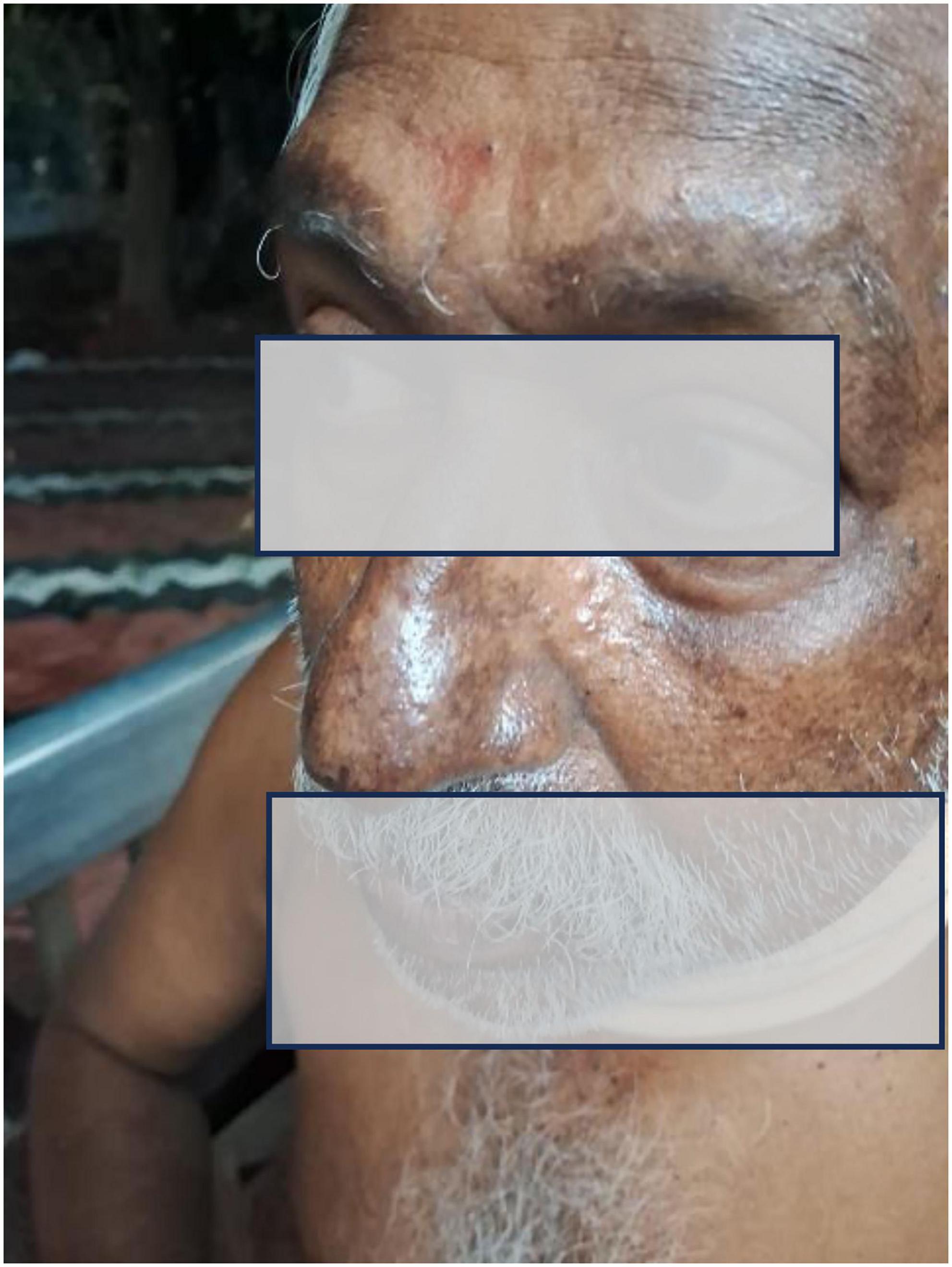
Figure 1. Clinical photograph demonstrating diffuse facial and periorbital hyperpigmentation with malar accentuation.
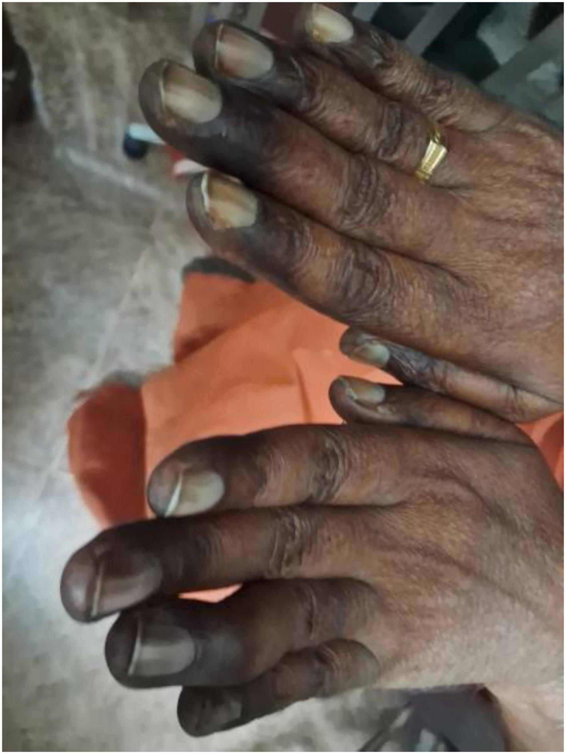
Figure 2. Bilateral dorsal hand hyperpigmentation, knuckle accentuation, and longitudinal melanonychia-like nailbed pigmentation.
No postural hypotension was detected. Serum cortisol was 18.6 μg/dL (reference 6.2–19.4 μg/dL), and electrolytes were normal, effectively excluding adrenocortical insufficiency as the cause of pigmentation.
Neurological examination revealed normal higher cognitive functions and intact cranial nerves, including fundoscopy (no optic atrophy or papilledema). Motor examination demonstrated normal bulk in all four limbs. Upper limb tone and power were normal (Medical Research Council [MRC] grade 5/5). In the lower limbs, mild bilateral spasticity was present with bilateral hip flexor weakness (MRC grade 4/5); remaining lower limb muscle groups were MRC 5/5. Deep tendon reflexes were symmetrically brisk at the knees and ankles; plantar responses were extensor bilaterally, indicating pyramidal tract involvement.
Sensory examination disclosed a dissociated pattern. Vibration sense was absent in the bilateral lower limbs to the hip level and impaired in the fingers of both hands, indicating extensive posterior column involvement. Joint position sense was impaired in the lower limbs. Pain and temperature sensation were relatively preserved, consistent with intact spinothalamic function. Romberg’s test was markedly positive. The “wash basin sign” (8) was positive. Gait was broad-based and sensory ataxic; tandem walking, significantly impaired. Heel-knee-shin testing showed mild proprioceptive impairment; finger-nose testing was normal. Neurological localization indicated bilateral posterior column and corticospinal tract involvement at the cervical cord level—the hallmark of SCD.
Timeline
For further details, please see Table 1.
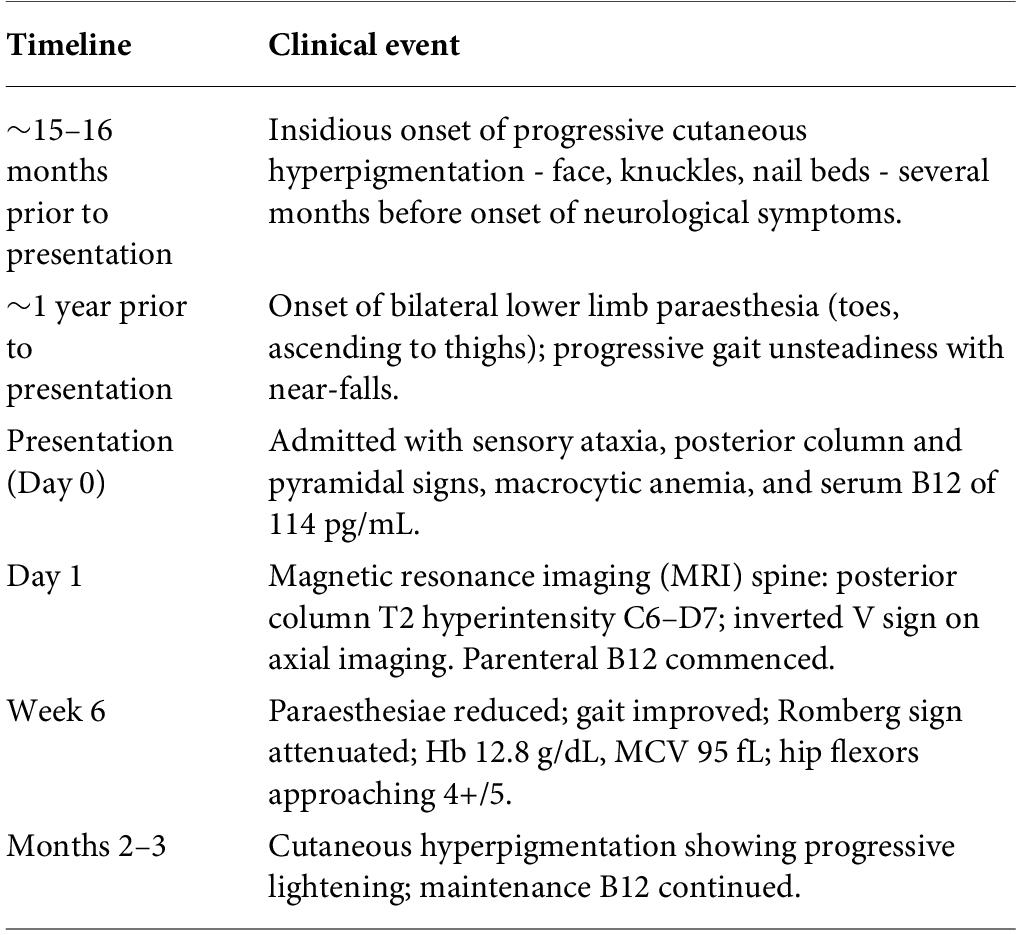
Table 1. Clinical timeline.
Diagnostic assessment
Hematological evaluation revealed macrocytic anemia: hemoglobin 10.7 g/dL, MCV 111.5 fL, red blood cell (RBC) 2.8 × 10⁶/μL, packed cell volume (PCV) 32%. Peripheral blood smear demonstrated macrocytes, oval macrocytes, hypersegmented neutrophils (>5% with ≥5 lobes), and anisocytosis, consistent with megaloblastic hematopoiesis. Serum vitamin B12 was markedly subnormal at 114 pg/mL (reference 200–900 pg/mL). Serum folate (12.4 ng/mL), serum copper (14.1 μmol/L), and serum cortisol (18.6 μg/dL) were all within normal limits. Full results are summarized in Table 2.
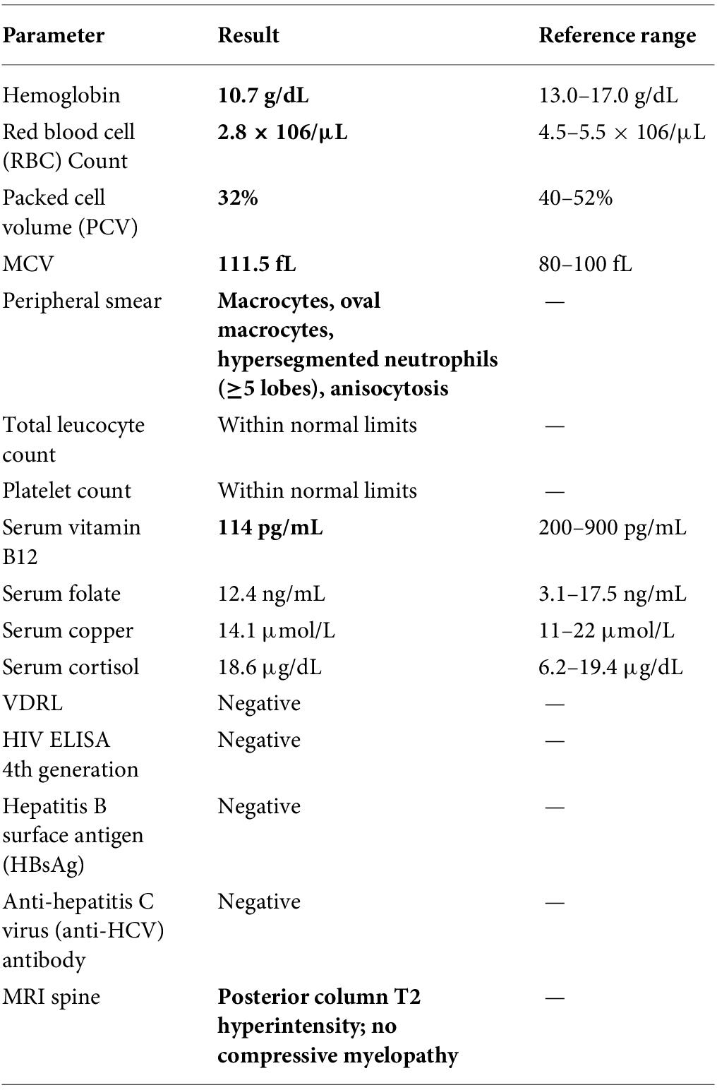
Table 2. Key investigations. Abnormal values shown in bold.
Serological screening was also performed: Venereal Disease Research Laboratory test (VDRL), HIV enzyme-linked immunosorbent assay (ELISA) (4th generation), hepatitis B surface antigen (HBsAg), and anti-hepatitis C virus (anti-HCV) antibody were all negative.
Magnetic resonance imaging (MRI) of the cervical and thoracic spine (Figures 3 and 4) demonstrated a long segment of T2 hyperintensity within the dorsal columns, beginning at C6 and extending to the D7 vertebral level on sagittal imaging. Axial T2-weighted images showed symmetric high signal confined to the dorsal columns, producing the characteristic inverted V sign—a radiological hallmark of SCD (9). There was no cord compression, intrinsic tumor, or demyelinating plaque to suggest an alternative etiology.
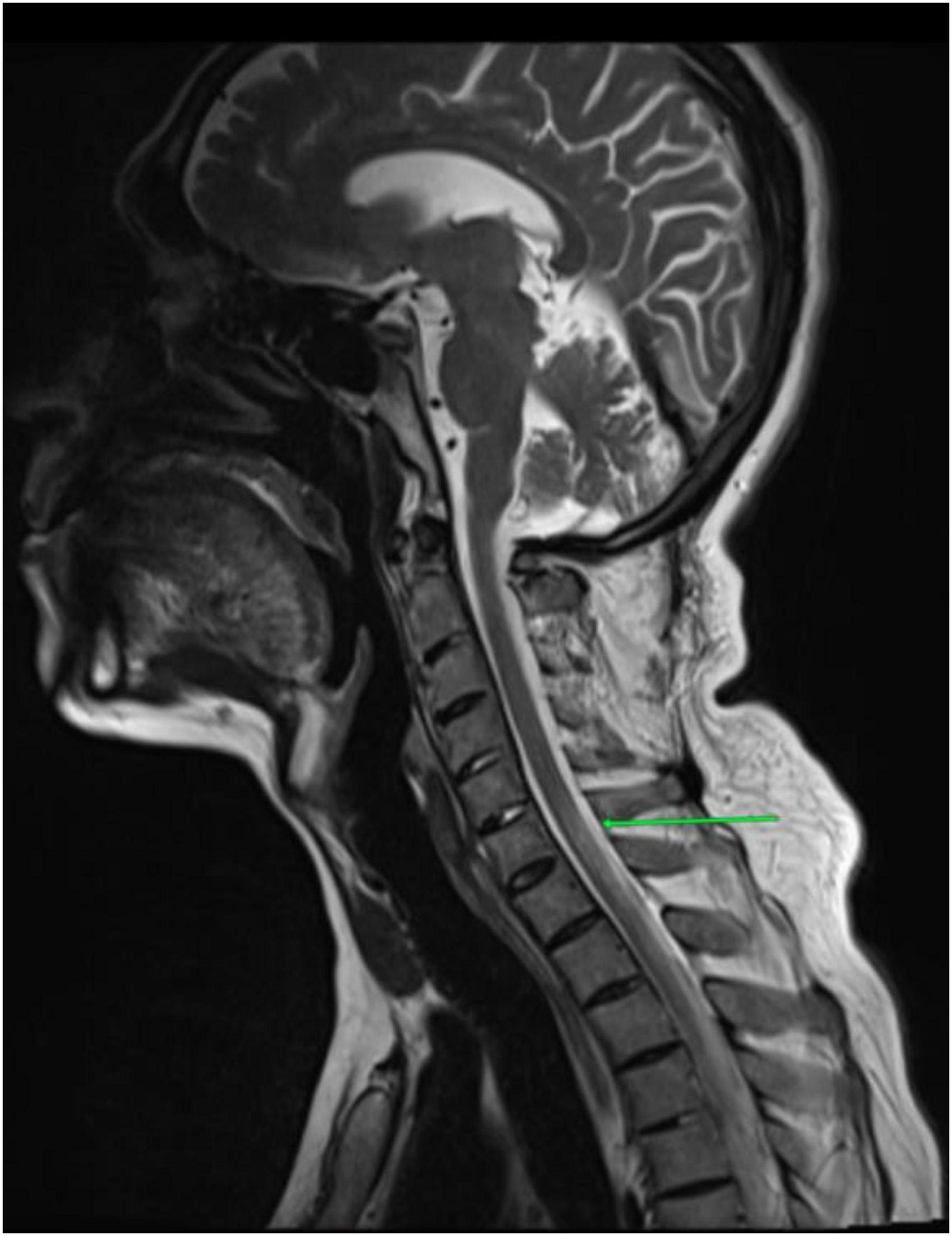
Figure 3. Sagittal T2-weighted MRI spine demonstrating long-segment posterior column T2 hyperintensity (green arrow) beginning at C6 and extending to D7.
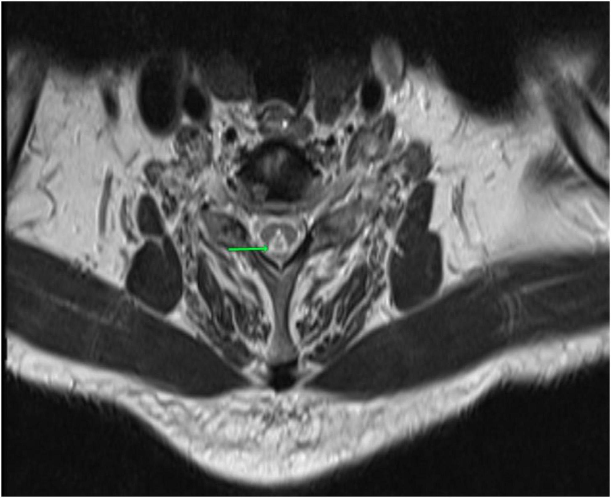
Figure 4. Axial T2-weighted MRI at the level of the cervical cord demonstrating bilateral symmetric dorsal column hyperintensity producing the inverted V sign (green arrow).
The following differential diagnoses were systematically evaluated. Copper deficiency myelopathy was excluded by normal serum copper (14.1 μmol/L). Compressive cervical myelopathy was excluded by MRI (important given rheumatoid arthritis (RA) predisposition to atlantoaxial instability). Multiple sclerosis was excluded by the longitudinal contiguous posterior column lesion pattern and absence of relapsing course. Nitrous oxide toxicity was excluded by history. Neuromyelitis optica spectrum disorder (NMOSD) was considered unlikely because the selective dorsal column T2 signal with inverted V sign is atypical for NMOSD; there was no optic neuritis, area postrema syndrome, or relapsing course. AQP4-IgG was not tested (limitation). MOG-antibody-associated disease (MOGAD) was considered unlikely as the imaging phenotype and monophasic course are atypical; MOG-IgG was not tested (limitation). Neurosyphilis (tabes dorsalis) was considered unlikely in this clinical context; serum VDRL was negative. HIV-associated vacuolar myelopathy was excluded because the HIV ELISA (4th generation) was negative; HBsAg and anti-HCV antibody were also negative. HTLV-1-associated myelopathy (HAM/TSP) was considered less likely given the complete metabolic explanation and clear treatment response favoring SCD; HTLV-1 serology was not performed, which is a limitation. Paraneoplastic myelopathy was considered unlikely because there were no constitutional features or known malignancy; paraneoplastic antibody panel was not performed (limitation). Vitamin E deficiency was considered unlikely because there was no fat malabsorption; serum vitamin E not measured (limitation). Serum methylmalonic acid (MMA) and homocysteine were not measured (limitation). The diagnosis was confirmed by the complete clinical, biochemical, radiological, and therapeutic response.
Therapeutic intervention
Parenteral vitamin B12 was commenced as hydroxocobalamin 1000 mcg intramuscularly daily for 7 days, followed by weekly injections for 1 month, then monthly maintenance (10). Folic acid supplementation was continued. The rheumatology team was consulted regarding the ongoing indication for MTX, balancing RA disease control against the documented neurological injury from cobalamin depletion.
Follow-up and outcomes
Progressive clinical improvement was evident within 6 weeks. Lower limb paraesthesiae reduced from constant bilateral distal tingling to occasional mild symptoms. Gait improved from a broad-based ataxic pattern requiring support to independent ambulation indoors; tandem walking partially recovered. Romberg’s sign attenuated from markedly positive to mildly positive, and no further near-falls were reported. Hip flexor strength showed partial recovery (MRC grade 4/5 to approaching 4+/5). Hematological assessment at 6 weeks demonstrated improvement in hemoglobin to 12.8 g/dL (from 10.7 g/dL at presentation) and MCV normalizing to 95 fL (from 111.5 fL). Cutaneous hyperpigmentation showed progressive lightening over subsequent months, consistent with restoration of S-adenosylmethionine (SAM)-mediated tyrosinase promoter methylation. Formal neurological outcome scales were not administered, as this was a routine clinical encounter; this represents a recognized limitation.
No adverse events related to treatment were observed.
Discussion
This case illustrates how MTX, prescribed for RA, can unmask latent cobalamin deficiency through a biochemical mechanism distinct from simple dietary lack. MTX inhibits dihydrofolate reductase, depleting methyltetrahydrofolate—the folate substrate for methionine synthase, which requires methylcobalamin as a co-factor (4). When MTX compromises this pathway, cobalamin-dependent methylation is further impaired beyond any underlying subclinical deficiency. Folic acid supplementation, while appropriate for hematological protection, does not rescue methionine synthase activity and may reduce the overt hematological signal that would otherwise prompt B12 testing (3).
The neurological injury in B12 deficiency arises from reduced synthesis of SAM, which is essential for myelin basic protein methylation (1, 2). Concurrent aberrant incorporation of methylmalonyl-CoA into myelin lipids compounds vacuolar myelopathy. The predilection for long fibers of the posterior columns and lateral corticospinal tracts explains the classical syndrome, and the extension of vibration loss to the fingers in this patient indicates advanced upward spread. The presence of a bilateral inverted V pattern on axial MRI—dorsal column signal selective, symmetric, and non-compressive—is the imaging signature of SCD and was clearly demonstrated in this case (9). Emerging evidence further suggests that SAM depletion in cobalamin deficiency may additionally impair SIRT1 (Sirtuin 1) activity—a NAD+-dependent deacetylase implicated in inflammatory regulation and cellular senescence—representing an area of potential mechanistic interest for future research.
The cutaneous phenotype — the true first symptom in this case — deserves particular emphasis. Facial, periorbital, knuckle, and nail bed hyperpigmentation resulting from B12 deficiency reflects reduced SAM-mediated methylation of the tyrosinase promoter, disinhibiting melanogenesis (6, 11). This pattern antedated neurological involvement by several months, making it the most accessible early clinical pointer and a potentially valuable screening trigger. The differential of Addison’s disease was excluded by normal serum cortisol and copper deficiency myelopathy by normal serum copper (12).
Strengths of this case include multimodal evidence—biochemical, radiological (characteristic inverted V sign on MRI), and a definitive therapeutic response—combined with systematic exclusion of mimics. VDRL, HIV ELISA, HBsAg, and anti-HCV were performed and were negative. Limitations include the absence of anti-intrinsic factor and anti-parietal cell antibody testing; serum MMA and total homocysteine not being measured (13, 14); AQP4-IgG, MOG-IgG, HTLV-1 serology, paraneoplastic antibody panel, and serum vitamin E not being performed; and formal neurological outcome scales not being administered. These reflect the inherent constraints of retrospective clinical case reporting in a resource-limited setting. The primary takeaway is threefold: (1) chronic MTX therapy is an under-recognized precipitant of SCD (15); (2) cutaneous hyperpigmentation is an early and clinically accessible warning sign preceding neurological involvement; and (3) folic acid supplementation cannot substitute for vigilant B12 surveillance in patients on antifolate therapy.
Patient consent
Written informed consent was obtained from the patient for publication of this case report and any accompanying images, including clinical photographs. The consent form is available upon request from the corresponding author.
Funding
No funding was received for this work.
Author contributions
KHM: clinical data collection, literature review, and manuscript drafting. MP: Neurological evaluation, MRI interpretation support, and manuscript critical revision. SR: clinical supervision and manuscript critical revision. All authors reviewed and approved the final manuscript.
Acknowledgments and AI disclosure
Artificial intelligence assistance was used only for language editing and formatting support. The authors reviewed, verified, and take full responsibility for all manuscript content.
Conflict of interest
The authors declare that the research was conducted in the absence of any commercial or financial relationships that could be construed as a potential conflict of interest.
References
2. Reynolds EH. The neurology of folic acid deficiency. In: Biller J, Ferro JM editors. Handbook of Clinical Neurology. (Vol. 120), Amsterdam: Elsevier (2014). p. 927–43.
3. Healton EB, Savage DG, Brust JC, Garrett TJ, Lindenbaum J. Neurologic aspects of cobalamin deficiency. Medicine (Baltimore). (1991) 70(4):229–45.
4. Weir DG, Scott JM. Brain function in the elderly: role of vitamin B12 and folate. Br Med Bull. (1999) 55(3):669–82.
5. Visser K, van der Heijde DM. Optimal dosage and route of administration of methotrexate in rheumatoid arthritis: a systematic review of the literature. Ann Rheum Dis. (2009) 68(7):1094–9.
6. Niiyama S, Mukai H. Reversible cutaneous hyperpigmentation and nails with white hair due to vitamin B12 deficiency. Eur J Dermatol. (2007) 17(6):551–2.
7. Gagnier JJ, Kienle G, Altman DG, Moher D, Sox H, Riley D , et al. The CARE guidelines: consensus-based clinical case reporting guideline development. J Med Case Rep. (2013) 7:223.
9. Ravina B, Loevner LA, Bank W. MR findings in subacute combined degeneration of the spinal cord: a case of reversible cervical myelopathy. AJR Am J Roentgenol. (2000) 174(3):863–5.
10. Langan RC, Goodbred AJ. Vitamin B12 deficiency: recognition and management. Am Fam Physician. (2017) 96(6):384–9.
11. Aaron S, Kumar S, Vijayan J, Jacob J, Alexander M, Gnanamuthu C. Clinical and laboratory features and response to treatment in patients presenting with vitamin B12 deficiency-related neurological syndromes. Neurol India. (2005) 53(1):55–8.
13. Patel AV, Morgan SL, Green R, Danila MI, Merriman TR, Wanzeck K , et al. Vitamin B12 status and hyperhomocysteinemia in patients with rheumatoid arthritis treated with methotrexate and folic acid. Am J Med Sci. (2024) 368(1):33–39.
14. Al-Jizani AS, Pathak S, Palit P, Achufusi N. Subacute combined degeneration of the spinal cord caused by an impairment in the functional vitamin B12 metabolic pathway. Cureus. (2024) 16(11):e73617.
15. Qudsiya Z, De Jesus O. Subacute combined degeneration of the spinal cord. In: StatPearls. Treasure Island (FL): StatPearls Publishing (2024). Available from: https://www.ncbi.nlm.nih.gov/books/NBK559316/ (Accessed March 20, 2024).
© The Author(s). 2026 Open Access This article is distributed under the terms of the Creative Commons Attribution 4.0 International License (https://creativecommons.org/licenses/by/4.0/), which permits unrestricted use, distribution, and reproduction in any medium, provided you give appropriate credit to the original author(s) and the source, provide a link to the Creative Commons license, and indicate if changes were made.