Introduction
Intracranial hypotension, also called Sagging brain syndrome, occurs due to alteration in flow, generation, and absorption of cerebrospinal fluid resulting in reduction of pressure in the cranial cavity. As a result, brain sagging occurs, which pulls supporting structures of brain. CSF leak is considered as the underlying etiology. Patients with headache that is orthostatic in nature showing improvement in lying posture with characteristic MRI findings should raise suspicion of Spontaneous Intracranial Hypotension (SIH).
Objective
1. To enumerate etiologies of SIH.
2. To describe the symptomatology and relevant investigations for SIH
3. To describe the newer treatment modalities of SIH
Clinical scenario
A 38-year-old lady with bronchial asthma, psychiatric illness, suicidal attempt by hanging 6 years ago presented with complaints of headache for more than 2 weeks. She was diagnosed with recurrent sinusitis and migraine and was on treatment at a local hospital and was referred to a higher center due to persistent headache associated with giddiness. Headache was gradual in onset, dull aching, intermittent in nature, predominantly over the occipital area and the frontal area, aggravated on erect posture and coughing, coughing and relieved on supine position. No h/o head injury, photophobia, vomiting, blurring of vision, altered sensorium, or double vision. On examination, the patient was conscious, oriented, and systemic examination was within normal limits.
MRI Brain showed Mucosal retention cyst in the maxillary sinus.
In view of persisting severe headache while getting up from bed and standing, the patient underwent MRI spine, which showed extensive extradural collection separating the anterior dural sac from the bony canal extending from C3 to D1 (Figure 1), following which CT Myelogram showed cervical dural leak anteriorly at C7-D1 (Figure 2) and Bony osteophyte impinging on dural sac – likely cause of leak (Figure 3).

Figure 1. MRI C spine sagittal T2 W sequence. Extensive extradural collection seen separating the anterior dural sac from the bony canal from the upper border of C3 to the lower border of D1.

Figure 2. PostCT myelogram sagittal image showing cervical dural leak emitting contrast into the anterior epidural space between the C7 and C8 levels.

Figure 3. PostCT myelogram axial image Bony osteophyte seen impinging on the anterior dural sac – likely cause of the dural leak.
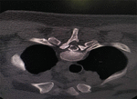
CT-guided blood patch done at C7-D1 level
Initially, the patient was managed with conservative measures including adequate hydration and bed rest with foot end elevation for 1 week. In view of persisting symptoms, Autologous Epidural Blood patch repair was carried out at C7-D1 epidural space via transforaminal route with 4 ml of autologous blood (Figure 4). The patient got symptomatically better within 2 days and hence discharged.
Etiology
CSF Leak is considered as the underlying cause of SIH (1). It can be congenital, traumatic, or iatrogenic. The major causes of CSF leak are dural membrane defect, leak from meningeal diverticulum, CSF-venous fistula, protrusion of osteophytes, disk herniation, and over drainage of CSF shunt (2). It can be associated with structural abnormalities in congenital connective tissue disorders. The triggering factors for CSF leak include fall, Valsalva maneuver, sneezing, sudden twist, sexual intercourse, sports, or trivial trauma (3).
Epidemiology
The estimated incidence is 5 in 1,00,000 per year (2), the peak age group being 40 years, predominantly affecting women (2). A categorization scheme was framed in classifying leaks in CSF flow according to etiology and association with extradural CSF collection on spine imaging into 4 types (4).
Type I CSF leak results from tear of dura mater (27% leaks), Type II from meningeal diverticula (42%), Type III from CSF-Venous Fistula (2.5%), and Type IV from indeterminate source (29%) (4). CSF leak mostly occurs at thoracic or cervicothoracic region.
Pathophysiology
CSF provides buoyancy to support the brain and spinal cord. As the CSF pressure reduces due to leak, buoyancy gets affected resulting in sagging of brain inside the cranium. It causes pull over the pain-sensitive parts of the brain causing headache, which gets aggravated in the upright position due to gravity (3). Headache can also occur due to cerebral vasodilation as a compensatory mechanism to low CSF pressure. The concept of CSF hypovolemia can be considered in patients with normal CSF pressures instead of CSF hypotension, though having typical clinical and radiological features of SIH (5).
Discussion
Spontaneous intracranial hypotension requires high clinical suspicion for diagnosis. The patient usually presents with characteristic headache that is orthostatic in nature with associated symptoms like vomiting, nausea, dizziness, tinnitus, disruption of sleep–wake cycles, progressive behavioral symptoms, and cognitive dysfunction (3).
Brain MRI using Gadolinium is considered as the sensitive test for SIH. Fluid collections in subdural space, Diffuse enhancement of pachymeninges (M.C), venous engorgement, brain sagging, and enlarged pituitary are some of characteristic findings in SIH.
Spinal MRI features of CSF leak are fluid collections in epidural space, disruption in dural sac, epidural venous engorgement, diverticula in meninges. Diagnostic lumbar puncture (LP) mostly shows an opening pressure of <60 mmH2O. Myelography either CT/MR using intrathecal contrast is recommended for finding site of leak (6).
The treatment options include:
Conservative therapy for mild to moderate symptoms of <2-week duration includes bed rest, analgesics, hydration, caffeine, high salt intake (6).
Lumbar epidural blood patch— Considered in patients with symptoms for >2 weeks, head history of or spine trauma, headache of severe intensity, history suggestive of any connective tissue disorder, symptoms not resolving after an initial 2-week trial of conservative treatment (6). It involves infusion of autologous blood, approximately 10–20 mL (up to 40 mL), into epidural space in lumbar region percutaneously (7). If no response after first patch, second EBP at an interval of 2–4 weeks can be done (6).
For persisting symptoms, Myelography using intrathecal contrast is used to explore the leak site and Targeted Epidural Blood Patch is preferred (8, 9).
For Refractory cases, Surgical repair (10, 11), Epidural Fibrin Glue (12, 13), or Endovascular Repair (10, 14) is performed:
If uncertain about the location of CSF leak, selections for therapy include Epidural continuous infusion of Saline (15) or Dextran and Repeat Imaging studies.
Conclusion
A high level of clinical suspicion is crucial for initiating relevant radiological investigations to aid in the prompt diagnosis and treatment of SIH. In majority of cases, symptoms get improved within a span of 2 weeks (16). In our case, apart from routine investigations, the patient underwent CT Myelography, which showed the site of CSF leak, following which Autologous Epidural blood patch repair was done as no improvement noted with trial of conservative measures. There is favorable prognosis for those whose symptoms resolve spontaneously or with conservative treatment. It can recur in around 10 percent of cases irrespective of treatment (7).
References
1. Upadhyaya P, Ailani JA. Review of spontaneous intracranial hypotension. Curr Neurol Neurosci Rep. (2019) 19:22.
2. Schievink W. Spontaneous spinal cerebrospinal fluid leaks and intracranial hypotension. JAMA. (2006) 295:2286–96.
3. Liaquat M, Jain S. Spontaneous intracranial hypotension: StatPearls [Internet]. Treasure Island, FL: StatPearls Publishing (2023).
4. Schievink W, Maya M, Jean-Pierre S, Nuño M, Prasad R, Moser FG. A classification system of spontaneous spinal CSF leaks. Neurology. (2016) 87:673–9.
5. Mokri B. Spontaneous cerebrospinal fluid leaks: From intracranial hypotension to cerebrospinal fluid hypovolemia–evolution of a concept. Mayo Clin Proc. (1999) 74:1113–23.
6. Cheema S, Anderson J, Angus-Leppan H, Armstrong P, Butteriss D, Carlton Jones L, et al. Multidisciplinary consensus guideline for the diagnosis and management of spontaneous intracranial hypotension. J Neurol Neurosurg Psychiatry. (2023) 94:835–43.
7. D’Antona L, Merchan MA, Vassiliou A, Watkins LD, Davagnanam I, Toma AK, et al. Clinical presentation, investigation findings, and treatment outcomes of spontaneous intracranial hypotension syndrome: a systematic review and meta-analysis. JAMA Neurol. (2021) 78:329–37.
9. Dobrocky T, Nicholson P, Häni L, Mordasini P, Krings T, Brinjikji W, et al. Spontaneous intracranial hypotension: searching for the CSF leak. Lancet Neurol. (2022) 21:369–80.
10. Callen A, Timpone V, Schwertner A, Zander D, Grassia F, Lennarson P, et al. Algorithmic multimodality approach to diagnosis and treatment of spinal CSF leak and venous fistula in patients with spontaneous intracranial hypotension. AJR Am J Roentgenol. (2022) 219:292–301.
11. Schievink W, Morreale V, Atkinson J, Meyer F, Piepgras D, Ebersold M. Surgical treatment of spontaneous spinal cerebrospinal fluid leaks. J Neurosurg. (1998) 88:243.
12. Mamlouk M, Shen P, Sedrak M, Dillon W. CT-guided fibrin glue occlusion of cerebrospinal fluid–venous fistulas. Radiology. (2021) 299:409–18.
13. Schievink W, Maya M, Moser F. Treatment of spontaneous intracranial hypotension with percutaneous placement of a fibrin sealant: report of four cases. J Neurosurg. (2004) 100:1098–100.
14. Brinjikji W, Savastano L, Atkinson J, Garza I, Farb R, Cutsforth-Gregory JKA. Novel endovascular therapy for CSF hypotension secondary to CSF-venous fistulas. AJNR Am J Neuroradiol. (2021) 42:882–7.
15. Muram S, Yavin D, DuPlessis S. Intrathecal saline infusion as an effective temporizing measure in the management of spontaneous intracranial hypotension. World Neurosurg. (2019) 125:37–41.
16. Rando T, Fishman R. Spontaneous intracranial hypotension: report of two cases and review of the literature. Neurology. (1992) 42:481–7.
© The Author(s). 2024 Open Access This article is distributed under the terms of the Creative Commons Attribution 4.0 International License (https://creativecommons.org/licenses/by/4.0/), which permits unrestricted use, distribution, and reproduction in any medium, provided you give appropriate credit to the original author(s) and the source, provide a link to the Creative Commons license, and indicate if changes were made.
