Introduction
A 60-year-old male presented to the outpatient department with a history of headache and seizures for the last 6 months. The symptoms were gradually progressive in course. She developed gait imbalance 2 months after the onset of the first symptoms. On examination, her GCS was 13/15 (E3V4M6). The patient underwent MRI, which revealed multiple cystic SOL in the subarachnoid space of the right temporo-parietal region and the mesencephalic cisterns. These cysts were hypointense on T1-weighted images and hyperintense on T2-weighted images, with no contrast enhancement and mass effect on the pons and medulla (Figure 1). The most probable differential diagnosis was cystic glioma, hydatid cyst, and dermoid cyst of the right temporal-parietal region (1, 2). The patient was taken for surgery following consent in May, 2024. The intra-op picture (Figure 2) shows the cyst being excised using the Dowling technique. The cyst was glistening white and avascular. As expected, it was multicystic, with delicate walls. Therefore, careful dissection was made to remove these cystic structures (3) (Video 1). These cysts were compressing the right temporal-parietal region. One by one, the cysts were removed, and upon observation during removal, it was found that the cysts contained sand-like particles, scolices; therefore, extra care was taken to avoid dissemination of these particles into the right temporal-parietal region (2, 4). Using normal saline irrigation, the maximum number of approachable cysts was removed (Figure 3) (Video 2). Surgery was uneventful, except for minimal ooze from the surgical site, which was controlled with Surgicel and gentle pressure. After closure, the patient was intubated and shifted to the surgical ICU for postoperative monitoring. On the second post op day, a CT scan of the brain was done, revealing a minimal surgical site bleed. Ischemic changes in the region of the right temporal-parietal were also observed. Gentle extubation was successful, and patient has recovered slowly. The (Figure 4) report confirmed the presence of a Hydatid cyst (2). A CT abdomen and pelvis was performed to diagnose any other cysts, and a second cyst was found in the liver (4, 5). The infectious disease team was consulted, and the patient was started on the recommended 400 mg BD for 1 month (6). Follow-up in the clinic after 10 Days revealed good healing of the wound edges; GCS 15/15, but persistent gait imbalance. CT Brain showed subtle improvement in mass effect, with persistence of gliotic changes within the right temporo-parietal region.
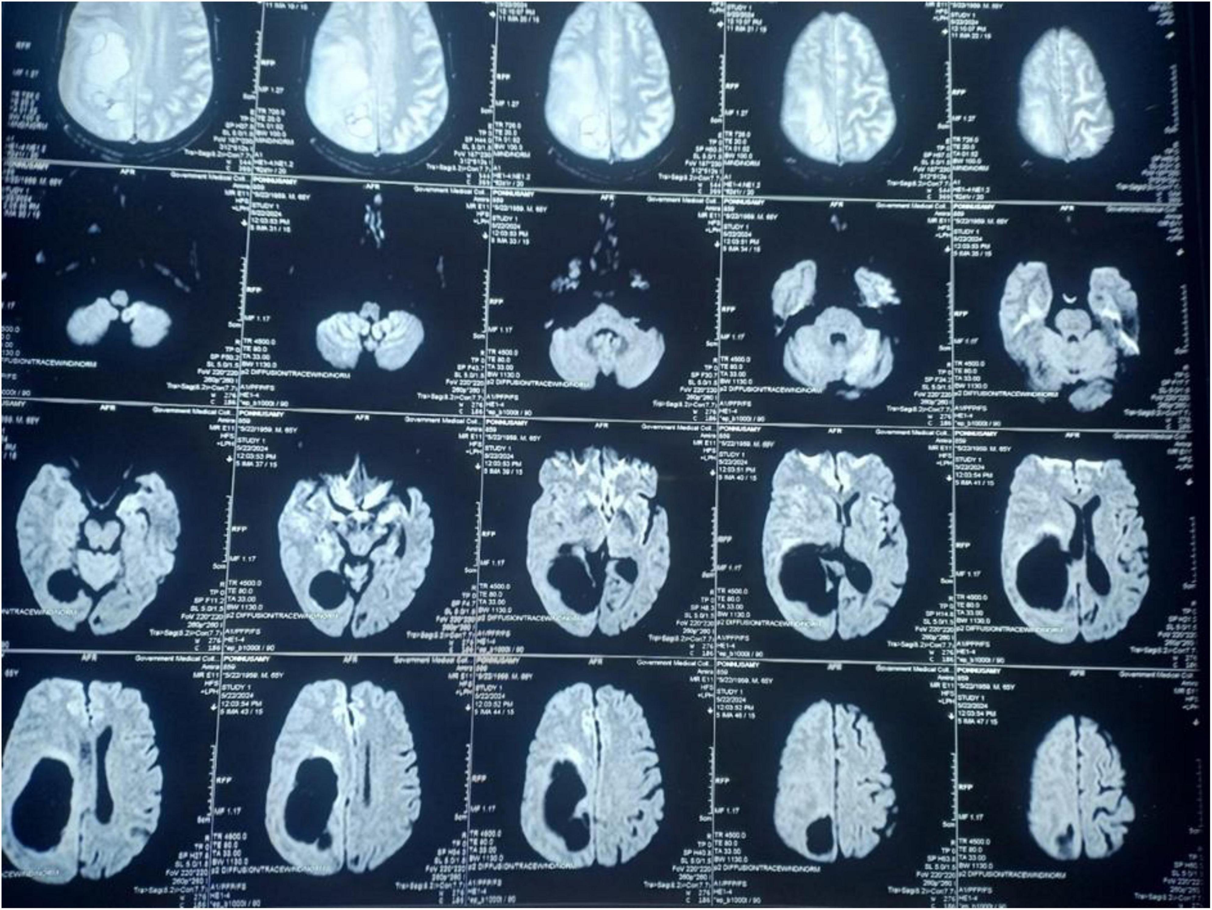
Figure 1. MRI Brain T2W images show multiple cystic space-occupying lesions.
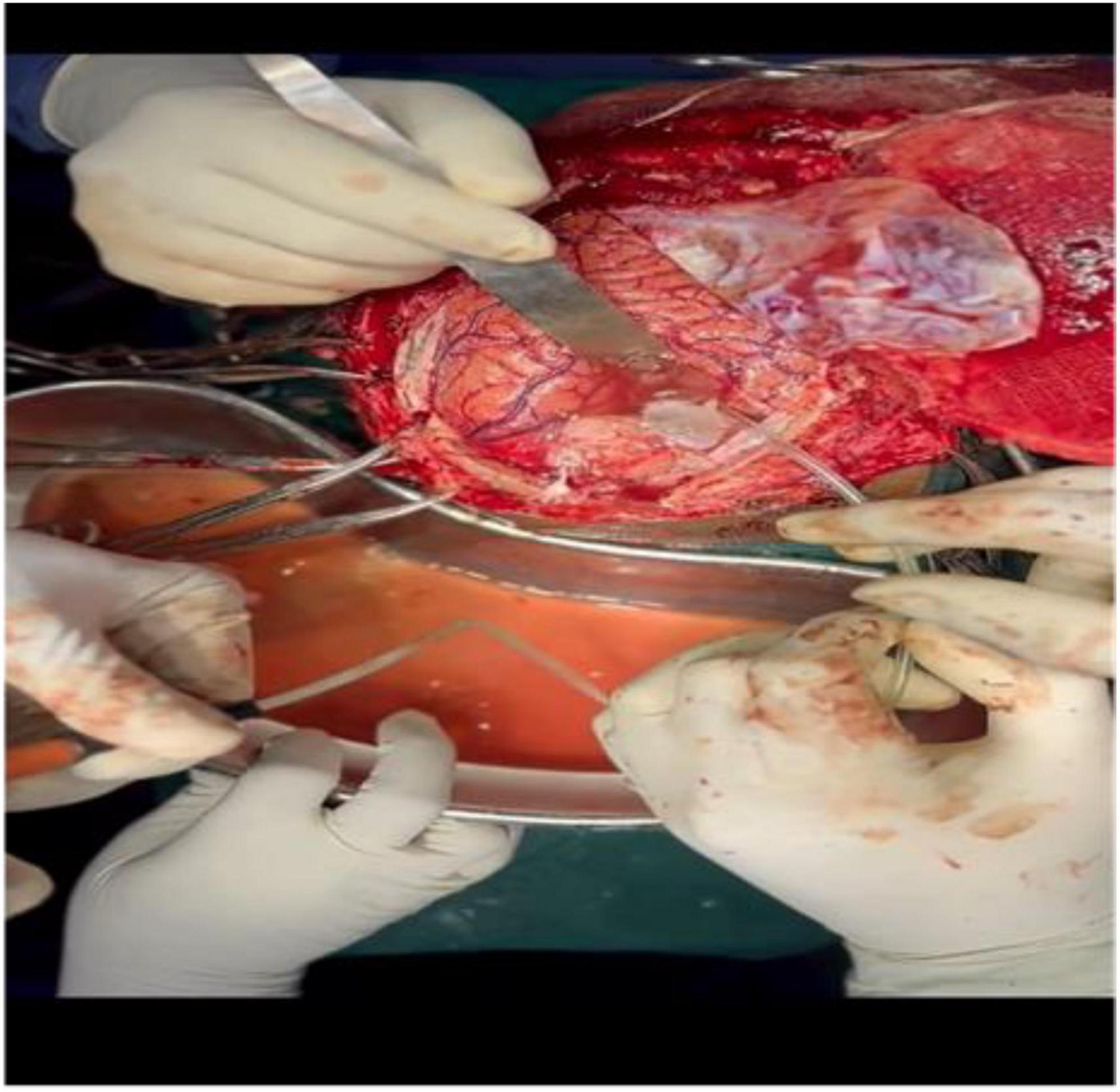
Figure 2. Intra op picture of the evacuation of a hydatid cyst using the dowling technique.
| Video 1. Application of the Dowling Technique. This video illustrates the careful preparation and application of the Dowling technique. The primary surgical goal is to deliver the cerebral hydatid cyst completely intact, as any rupture would cause harmful cystic fluid to spill into the brain parenchyma. https://youtube.com/shorts/K0Dr0hr1L28 |
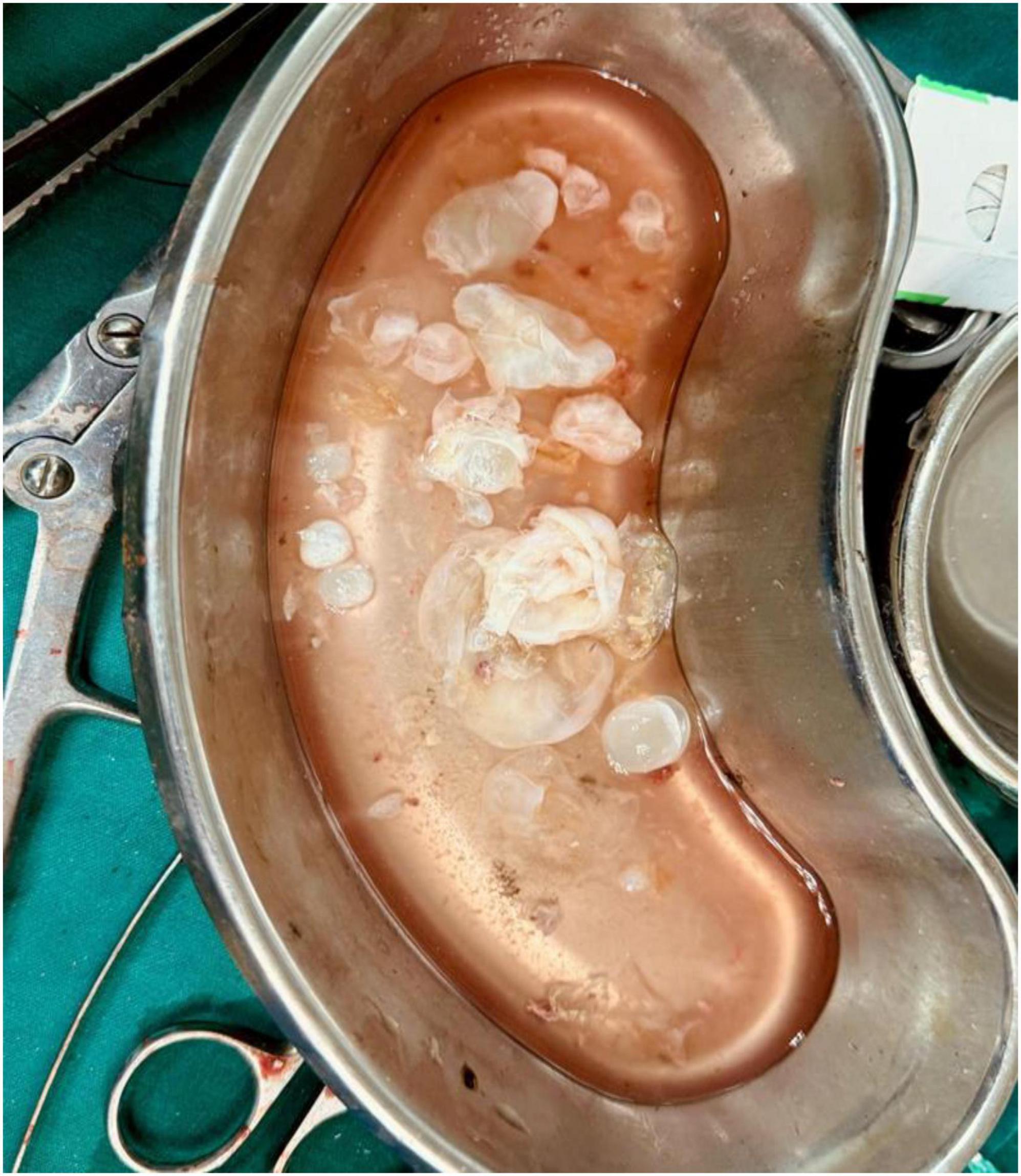
Figure 3. Multiple hydatid cysts, 20–25 in number.
| Video 2. Critical Intact Extraction. This video captures the high-stakes final moments of the cyst’s extraction. https://youtube.com/shorts/8R7fotwJfKA |
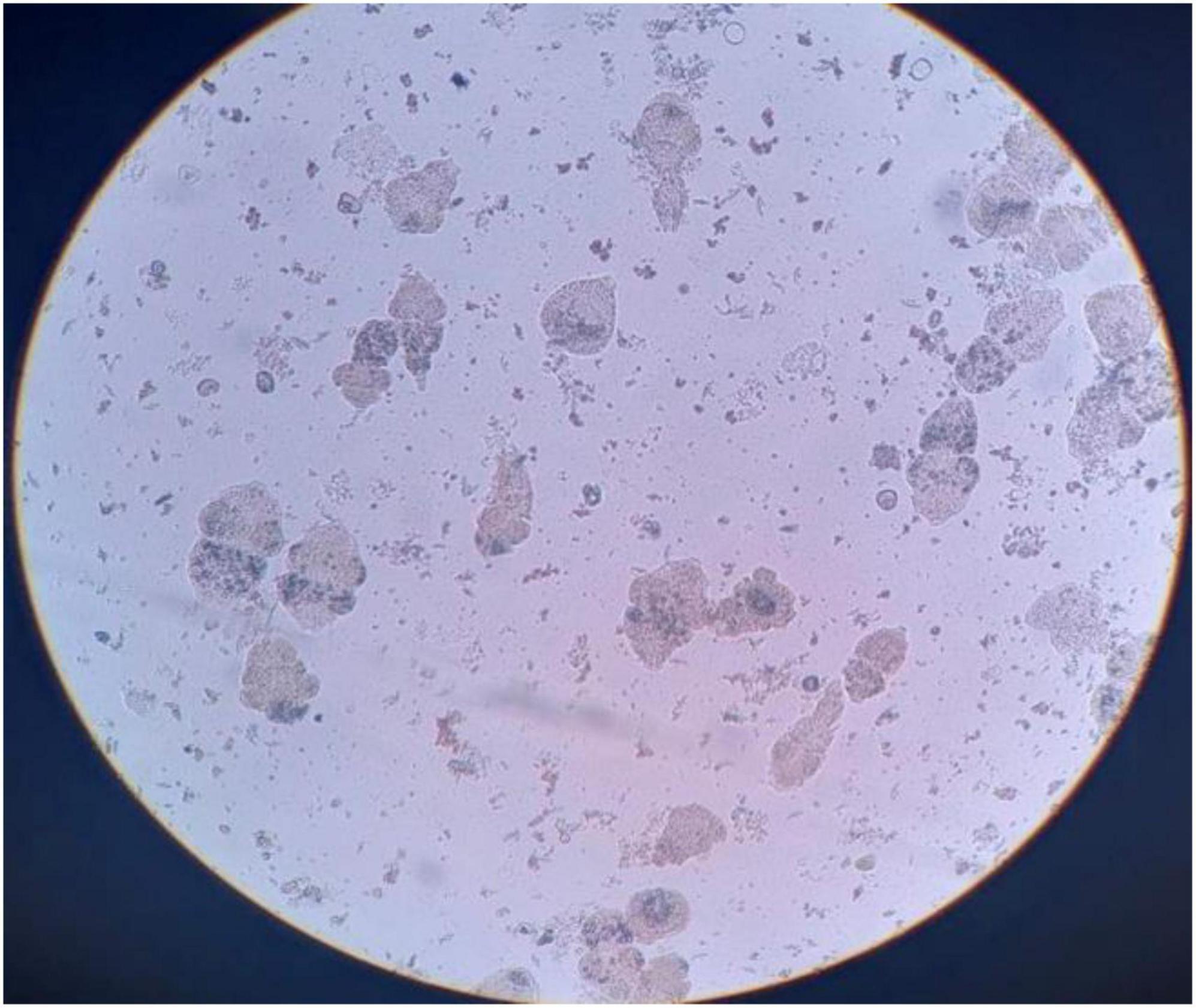
Figure 4. Microscopic picture of a hydatid cyst.
Discussion
Hydatid cyst is classified as a zoonosis caused by Echinococcus granulosus (2, 4). The dog is the definitive host, which harbors the disease by eating infected tissues from sheep, goats, and other animals (4). Contaminated food is accidentally consumed by humans, who serve as intermediate hosts. In humans, the larva enters the blood and reaches different organs, especially those with a rich blood supply (1, 4). While CNS spread is considered rare, it is the third most common site of involvement (5). The following is a list of organs where hydatid cysts can spread: muscles, the orbit, the heart, the adrenal gland, the thyroid, the submandibular gland, the kidneys, the intestine, the internal acoustic canal, the jugular foramen, and the spine (4, 5). Radiologically, Hydatid cysts are best diagnosed on contrast-enhanced MRI (1). The cysts can be solitary or multiple. The clinical signs and symptoms depend on the area of brain involvement and range from Headache to focal neurological deficits in the affected area when the cyst is present (1, 2). Histopathological diagnosis of hydatid cyst is confirmatory by observing scolices (the sand) in the Cystic fluid (2). During surgery, Saline (Dowling technique) is used to dissect the hydatid cyst, and with this method, the cysts are usually delivered intact (3). Microscopic dissection is done with care to avoid the spillage of cyst contents in the surrounding areas (3). Once the diagnosis of hydatid disease is confirmed, anti-parasitic treatment Albendazole is given 400 mg orally BID for 1–3 months (6). Reviewing the literature from 2008 to 2018 using the PubMed search engine, we found that the disease is geographically concentrated in the Middle East and developing countries (5).
Funding
The authors declare that no financial support or funding was received for this study.
Conflict of interest
The authors declare that the research was conducted in the absence of any commercial or financial relationships that could be construed as a potential conflict of interest.
References
1. Winn HR editor. Youmans and Winn Neurological Surgery. 7th ed. Philadelphia, PA: Elsevier (2017).
2. Kumar V, Abbas AK, Fausto N, Mitchell RN. Robbins Basic Pathology. 8th ed. Philadelphia, PA: Saunders/Elsevier (2007).
3. Schmidek HH, Roberts DW editors. Schmidek and Sweet Operative Neurosurgical Techniques: Indications, Methods, and Results. 5th ed. Philadelphia, PA: Saunders/Elsevier (2005).
4. Paniker CKJ, Ghosh S. Paniker’s Textbook of Medical Parasitology. New Delhi: Jaypee Brothers Medical Publishers (2018).
5. Gupta S, Desai K, Goel A. . Intracranial hydatid cyst: a report of five cases and review of literature. Neurol India. (1999) 47(3):214–7.
6. Ramamurthi R, Tandon PN, Behari S, Vasudevan MC, Kumar G et al. Ramamurthi and Tandon’s Textbook of Neurosurgery. 4th ed. New Delhi: Jaypee Brothers Medical Publishers (2026).
© The Author(s). 2026 Open Access This article is distributed under the terms of the Creative Commons Attribution 4.0 International License (https://creativecommons.org/licenses/by/4.0/), which permits unrestricted use, distribution, and reproduction in any medium, provided you give appropriate credit to the original author(s) and the source, provide a link to the Creative Commons license, and indicate if changes were made.