Introduction
Atypical teratoid/rhabdoid tumor (ATRT) is a highly malignant embryonal tumor of the central nervous system that occurs predominantly in children under 3 years of age. It is defined by biallelic inactivation of SMARCB1 (INI1) and, in a small minority of cases, SMARCA4 and is classified as a Central Nervous System World Health Organization (CNS WHO) grade 4 neoplasm (1, 2). Roughly half of all cases arise in the posterior fossa, where the cerebellar hemisphere and the cerebellopontine angle are recognized sites of origin (2, 3).
The radiological appearance of ATRT overlaps substantially with that of medulloblastoma and ependymoma. A large, heterogeneous, solid-cystic mass with intratumoral hemorrhage, calcification, and marked diffusion restriction in a child under 3 years should raise the possibility of ATRT, but the diagnosis remains histological and molecular (4). When such a lesion presents as an extra-axial cerebellopontine angle mass, the preoperative differential broadens further to include extra-axial medulloblastoma, laterally extending ependymoma, choroid plexus tumors, and uncommonly at this age, nerve sheath tumors (5).
Surgery in this setting is challenging on several counts. The tumor is frequently large relative to the volume of the infant’s posterior fossa, it is characteristically vascular and friable, and the cranial nerves of the cerebellopontine angle are typically thinned and splayed across its capsule. Blood loss that would be inconsequential in an adult is significant in an 11-month-old child. At the same time, the extent of resection is an established prognostic variable in ATRT, so the operation must aim at maximal safe removal without accepting a neurological or vascular injury to achieve it (2, 3, 6). In this operative video we demonstrate the excision of a giant right cerebellopontine angle ATRT in an infant through a suboccipital craniotomy extended to the right, undertaken 1 week after cerebrospinal fluid diversion had been performed in the emergency setting.
| Video 1. Microsurgical excision of a giant right cerebellopontine angle atypical teratoid/rhabdoid tumor in an 11-month-old infant through a suboccipital craniotomy extended to the right. The video demonstrates cerebrospinal fluid release and cerebellar relaxation, identification and preservation of the arachnoid plane, cauterization of the tumor feeders with preservation of the cerebellar and brainstem supply, intratumoral decompression, protection of the fourth ventricle with a cottonoid, dissection of the tumor from the brainstem and the cranial nerves of the cerebellopontine angle, and watertight closure. https://youtu.be/fgvFF21W9BI |
Clinical presentation
An 11-month-old male infant, born at term by spontaneous vaginal delivery and appropriate for gestational age, with an uneventful antenatal and postnatal course and no previous hospitalization, presented with 1 month of recurrent projectile, non-bilious, non-blood-stained vomiting containing food particles, associated with regurgitation of feeds through the nose. For 2 weeks before the presentation, there had been loss of previously acquired milestones, including head control, sitting, and crawling, together with deviation of the angle of the mouth to the left and an inability to close the right eyelid completely. There was no history of trauma, fever, seizures, altered sensorium, limb weakness, or limitation of activity. The child had been developmentally normal until the onset of the present illness.
On examination, the child was alert and active, with pupils 3 mm, equal and reactive to light. Cranial nerve examination demonstrated a right lower motor neuron facial palsy. Motor examination showed normal bulk and tone with power greater than 3/5 in all four limbs and reflexes graded 2+; the plantar responses were extensor bilaterally, which is physiological at this age. Sensations couldn’t be reliably examined. Cerebellar signs and signs of meningeal irritation were absent.
Non-contrast computed tomography of the brain demonstrated a heterogeneous, hyperdense solid-cystic lesion in the right cerebellopontine angle with mass effect on the brainstem, compression of the fourth ventricle, and obstructive hydrocephalus. Magnetic resonance imaging with contrast demonstrated a large 4 × 4 cm mass lesion in the right cerebellopontine angle, hypointense on T1-weighted and hypo- to iso-intense on T2/FLAIR sequences, with solid and cystic components, significant diffusion restriction, and enhancement of the solid component. There was brainstem and fourth ventricular compression with surrounding oedema and obstructive hydrocephalus (Figure 1). The lesion appeared extra-axial radiologically, and the radiological impression favored an extra-axial medulloblastoma. A comprehensive malignancy workup showed no evidence of another primary lesion or of metastatic disease. The child was admitted through the emergency department with intractable vomiting and features of raised intracranial pressure, and cerebrospinal fluid diversion was carried out at that time; definitive tumor surgery was undertaken 1 week later.
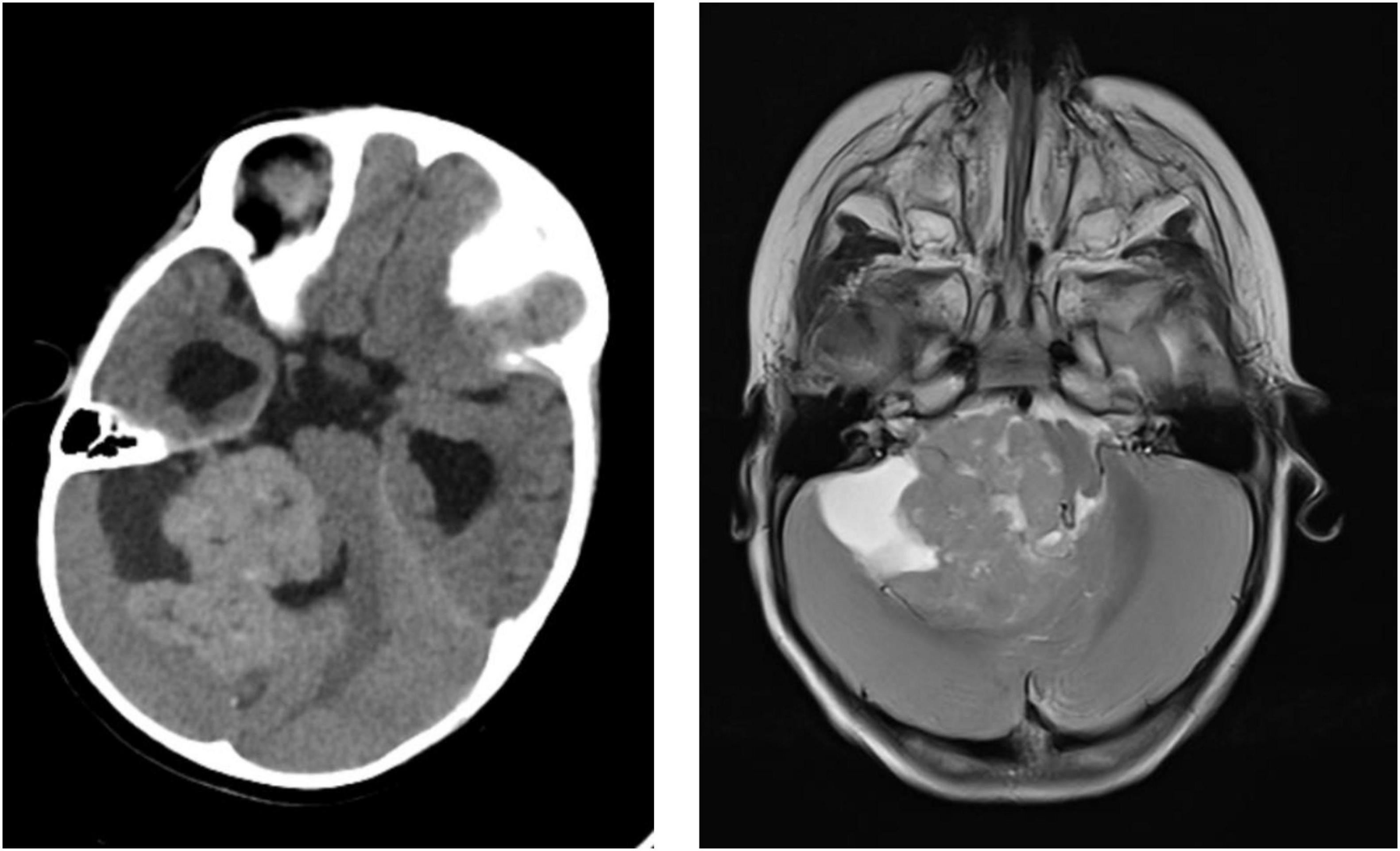
Figure 1. Preoperative imaging. Left: axial non-contrast computed tomography demonstrating a heterogeneous, hyperdense solid-cystic lesion in the right cerebellopontine angle with mass effect on the brainstem and fourth ventricle. Right: axial magnetic resonance image demonstrating the T2 hypo- to iso-intense extra-axial right cerebellopontine angle mass with solid and cystic components, brainstem and fourth ventricular compression, and obstructive hydrocephalus.
Operative technique
Surgical planning
Two objectives were defined preoperatively: relief of the obstructive hydrocephalus and maximal safe resection of the lesion with preservation of the brainstem and the neurovascular contents of the cerebellopontine angle. Cerebrospinal fluid diversion was carried out first, in the emergency setting, so that the posterior fossa would be decompressed before the dura was opened and the resection would not be performed against raised intracranial pressure. The interval that this bought was used deliberately. Tumor excision was deferred for 1 week while the dehydration produced by a month of projectile vomiting was corrected and a lower respiratory infection was treated so that a long, vascular posterior fossa resection was not undertaken in an infant who was volume-depleted and respiratorily compromised. The lesion filled the whole of the right cerebellopontine angle and reached the midline structures, and a suboccipital craniotomy sited on the midline but carried predominantly to the right, as far as the mastoid tip, gave a direct trajectory to the angle together with control of the fourth ventricle, the cisterna magna, and the foramen magnum.
Cerebrospinal fluid diversion and preoperative optimization
A ventriculoperitoneal shunt was inserted through a left Frazier’s point burr hole in the emergency setting, in view of intractable vomiting and obstructive hydrocephalus. The contralateral side was chosen deliberately so that the burr hole and the subcutaneous tract lay well away from the planned posterior fossa incision. Drainage was allowed to proceed gradually; abrupt decompression of a dilated ventricular system in the presence of a large posterior fossa mass risks upward transtentorial shift, and slow, controlled release avoids this. The distal catheter was tunneled, and the abdominal end was secured. Tumor excision was then deliberately deferred for 1 week. Over that interval the vomiting settled, hydration and electrolytes were corrected, and a lower respiratory infection was treated, so that the child came to the definitive operation in a considerably better physiological state than at presentation.
Positioning and exposure
For the definitive procedure the child was placed in the lateral position. The head was supported and secured with adhesive tape rather than with pin fixation, which is unsuitable for the thin infant skull, with the neck slightly flexed and all pressure points padded. Normothermia was maintained actively, and secure wide-bore venous access was obtained in anticipation of blood loss from a vascular tumor. A hockey-stick incision was marked: it began in the midline above the inion, curved laterally towards the mastoid tip, and extended inferiorly to the level of the second cervical vertebra (Figure 2). The suboccipital muscles were split and reflected together with the scalp flap. A suboccipital craniotomy was then carried out, extending approximately 2 cm to the left of the midline and 4 cm to the right, laterally as far as the mastoid tip and inferiorly to the rim of the foramen magnum. This asymmetrical, predominantly right-sided exposure gave a flat working trajectory into the cerebellopontine angle while retaining access to the midline and minimized the need for cerebellar retraction.
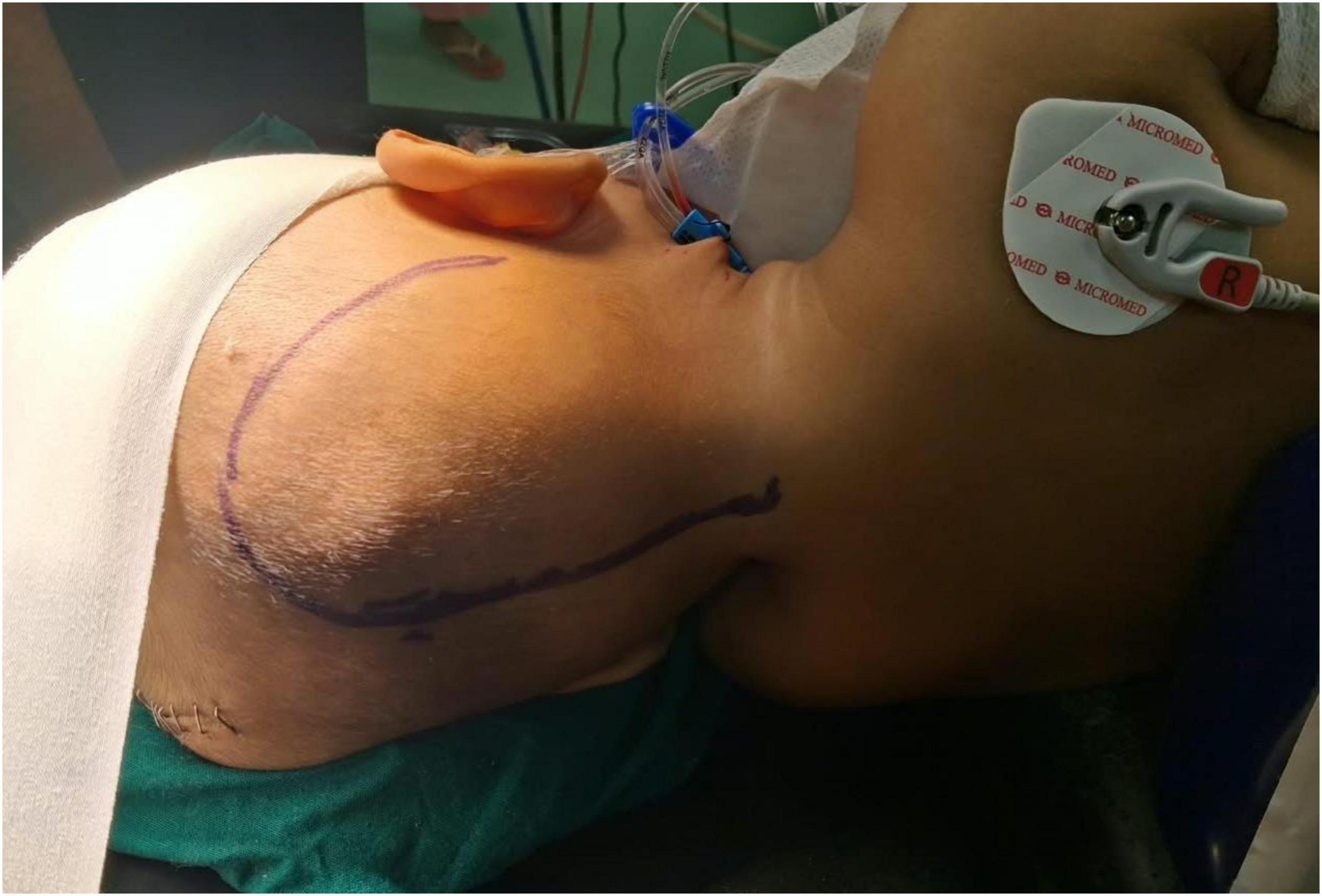
Figure 2. Positioning and skin incision. The child is in the lateral position with the head supported and secured with adhesive tape rather than pin fixation. The hockey-stick incision is marked: it begins in the midline above the inion, curves laterally towards the mastoid tip, and extends inferiorly to the level of the second cervical vertebra.
Dural opening and cerebellar relaxation
A cruciate dural opening was fashioned and tacked up. The cisterna magna and the cerebellomedullary cistern were opened and cerebrospinal fluid released slowly, which relaxed the cerebellum and opened the lateral corridor. Gravity-assisted retraction was relied upon throughout, since the immature cerebellar cortex tolerates fixed retraction poorly. The operating microscope was introduced at this stage, and the operative video begins at this transition.
Tumor exposure and the arachnoid plane
A yellowish-gray, soft, suckable, and highly vascular tumor with a few hard areas, possibly representing calcification, was encountered. Contrary to the preoperative radiological impression, the lesion was not purely extra-axial. It infiltrated the lower lobe of the right cerebellar hemisphere and the tonsil and appeared to arise from the cerebellum itself, while filling the entire cerebellopontine angle and distorting and rotating the brainstem. A relatively well-maintained arachnoid plane was nevertheless present between the tumor and the neurovascular structures of the right cerebellopontine angle. The cranial nerves within the angle were thinned out and involved by the tumor and were identified and traced along the capsule before any debulking was undertaken. The tumor was cystic in certain areas. Establishing the arachnoid interface before entering the lesion is the single most useful maneuvre of the operation, because that plane remains the guide for every subsequent step.
Feeder cauterisation and intratumoral decompression
The feeding vessels of the tumor were identified and cauterized at the outset, with deliberate preservation of the vessels supplying the cerebellum and the brainstem; devascularizing the lesion while sparing the normal supply is what makes the subsequent debulking bloodless rather than merely faster. The capsular surface was coagulated with low-power bipolar cautery before it was entered. The cystic components were decompressed early, which immediately yielded working space in a confined posterior fossa. Intratumoral decompression was then performed well inside the capsule using suction and ultrasonic aspiration, with the arachnoid plane preserved throughout. Piecemeal removal from within was preferred: the tumor was friable and highly vascular, and any attempt to deliver it as a mass would have risked both avulsion of the capsule from the brainstem and blood loss that is poorly tolerated at this age.
Dissection from the brainstem and cranial nerves
Once the lesion had been sufficiently decompressed, the capsule was progressively delivered away from the brainstem into the resection cavity. Medially, the fourth ventricle was identified, and a cottonoid was placed within it before the medial dissection was carried further; blood spilled into the fourth ventricle may pass through the aqueduct into the lateral ventricles, and protecting the ventricle at this point avoids that. Anteriorly, the brainstem was identified and followed as the deep limit of the dissection. A plane was preserved between the tumor, the brainstem, and the adjacent cerebellum throughout. Sharp dissection under high magnification was used at the interface with the cranial nerves, and traction on the nerves was avoided. Perforating vessels running to the brainstem were traced and preserved rather than coagulated, since the consequences of sacrificing a perforator in this region are disproportionate to any gain in speed.
Extent of resection and closure
Near total excision of the tumor was achieved. A thin layer of tumor adherent to the brainstem, together with a small portion adherent to the cranial nerves, was deliberately left behind in order to avoid brainstem infarction and neurovascular injury. Haemostasis was secured with irrigation, low-power bipolar coagulation, and topical haemostatic agents, and the cottonoid protecting the fourth ventricle was removed only once the field was dry. A watertight duroplasty was performed. The bone was replaced and secured with Prolene sutures, and the wound was closed in layers.
Surgical nuances and technical pearls
First, cerebrospinal fluid diversion before the posterior fossa is opened converts the operation. Treating the hydrocephalus first delivers a relaxed cerebellum and a workable corridor and removes the need to operate against raised intracranial pressure in a child whose intracranial compliance is already exhausted. Drainage should nonetheless be gradual, and the shunt entry point should be sited away from the planned posterior fossa incision. Equally important is what the shunt buys: an interval in which a dehydrated, vomiting infant with an intercurrent chest infection can be corrected. In this case the tumor operation was deliberately deferred by a week for that purpose, and the child came to surgery in a far better physiological state.
Second, the arachnoid plane should be defined before the tumor is entered, not after. In this case the plane between the tumor and the neurovascular structures of the cerebellopontine angle was relatively well maintained despite the size of the lesion. Identifying it at the outset, and returning to it repeatedly, is what allows a large tumor to be removed without a cranial nerve or brainstem injury. Once the plane is lost, it is rarely recovered.
Third, the sequence of feeder cauterization, cyst decompression, and intratumoral debulking should precede any attempt at circumferential mobilization. Identifying the feeding vessels of the tumor and coagulating them, while preserving the vessels that supply the cerebellum and the brainstem, limits blood loss; draining the cysts first creates space; and debulking from within converts a large fixed mass into a mobile shell that can be delivered away from the brainstem rather than pulled off it.
Fourth, the decision to leave a thin adherent layer of tumor is a legitimate operative judgment rather than a failure; the pursuit of complete resection at the skull base is justified only when the attendant long-term morbidity is minimal (7). Extent of resection carries prognostic weight in ATRT (6), but the remnant in this case lay on the brainstem and on cranial nerves that were already thinned by the tumor, and the lesion was planned for adjuvant therapy. In an 11-month-old child, a thin residue is preferable to a brainstem infarct or a lower cranial nerve palsy.
Fifth, the fourth ventricle should be protected before the medial dissection is carried further. Once the ventricle is opened medially, a cottonoid placed within it keeps blood out of the ventricular system; blood that reaches the fourth ventricle can pass through the aqueduct into the lateral ventricles, and in a child who already has a shunt in situ, that is a complication worth a few seconds of prevention.
Sixth, posterior fossa closure in infancy deserves the same care as the resection. Watertight duroplasty and replacement of the bone protect against cerebrospinal fluid leak and pseudomeningocele, complications that are common after posterior fossa surgery in this age group and that can delay the initiation of adjuvant treatment.
Postoperative outcome and pathological findings
There were no intraoperative or postoperative complications. The child was transferred to the pediatric intensive care unit for monitoring, was extubated after 3 hours, and was weaned to room air over the subsequent 18 hours. Nasogastric feeds were commenced on the first postoperative day and were well tolerated. In view of the clinical improvement, haemodynamic stability and absence of features of raised intracranial pressure, the child was transferred to the ward for further management. Sutures were removed on the 14th postoperative day. At discharge the child was active, afebrile, and playful; the surgical site was healthy; and the right lower motor neuron facial palsy persisted.
Contrast-enhanced MRI of the brain with screening of the spine, performed 6 weeks after surgery, showed postoperative changes with a cerebrospinal fluid-containing cavity at the operative site. An ill-defined, diffusion-restricting, heterogeneously enhancing residual tumor, up to approximately 8 mm in thickness, lined the margins of the cavity and extended into the right cerebellopontine angle and the cerebellomedullary cistern and along the right middle and inferior cerebellar peduncles, adjacent to the brainstem and cranial nerves (Figure 3). An evolving subacute hematoma measuring approximately 2.1 cm lay along the inferior aspect of the cavity. A separate 8 × 9 × 8 mm diffusion-restricting, non-enhancing lesion in the right tectal plate/inferior colliculus was suspicious for an additional tumor deposit. Thin bilateral subdural collections with smooth dural enhancement were attributed to recent surgery and cerebrospinal fluid shunting. There was no hydrocephalus, and screening of the spine showed no leptomeningeal dissemination.
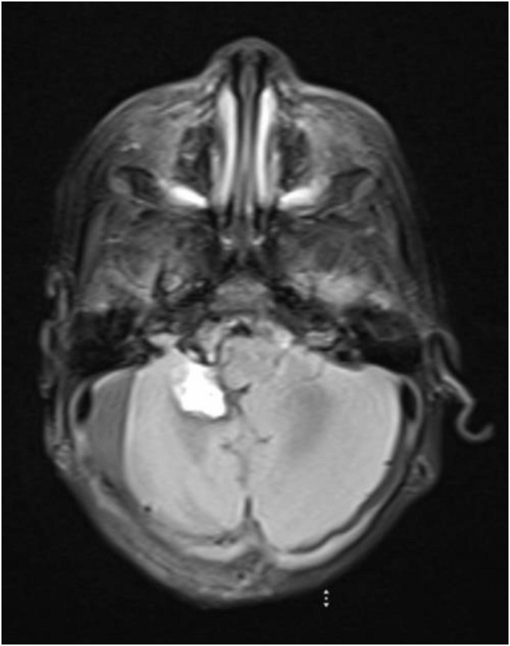
Figure 3. Postoperative axial T2-weighted magnetic resonance image at 6 weeks, showing the operative cavity in the right cerebellar hemisphere and cerebellopontine angle, with an evolving subacute hematoma along its inferior aspect and ill-defined residual tumor along the cavity margins adjacent to the brainstem.
Histopathological examination established a diagnosis of CNS WHO grade 4 ATRT. The diagnosis was not anticipated radiologically: the preoperative impression had favored an extra-axial medulloblastoma, which underlines the point that ATRT cannot be excluded on imaging grounds in an infant with a large solid-cystic posterior fossa mass showing marked diffusion restriction (4). The child was referred to medical oncology for adjuvant therapy, and surveillance magnetic resonance imaging was scheduled.
Conclusion
A giant cerebellopontine angle ATRT in an infant can be excised safely through a suboccipital craniotomy extended to the right when the operation is staged deliberately. Cerebrospinal fluid diversion in the first instance, with definitive surgery deferred until dehydration and an intercurrent respiratory infection have been corrected; early delineation and preservation of the arachnoid plane; cauterisation of the tumor feeders with preservation of the cerebellar and brainstem supply; protection of the fourth ventricle with a cottonoid; and intratumoral decompression before circumferential mobilization are the maneuvres that make the resection controlled rather than reactive. Leaving a thin layer of tumor on the brainstem and cranial nerves is an appropriate trade-off in this age group, particularly when adjuvant therapy is planned. The accompanying video illustrates these steps and may serve as a guide for the management of similar lesions.
Funding
The author(s) declared that financial support was not received for this work and/or its publication.
Consent
Written informed consent for the surgical procedure, for intraoperative video recording and for academic publication of the video and the accompanying clinical data was obtained from the parents of the child. All patient identifiers have been removed from the video and from the manuscript.
Conflict of interest
The authors declare that the research was conducted in the absence of any commercial or financial relationships that could be construed as a potential conflict of interest.
References
1. Louis DN, Perry A, Wesseling P, Brat DJ, Cree IA, Figarella-Branger D , et al. The 2021 WHO classification of tumors of the central nervous system: a summary. Neuro Oncol. (2021) 23(8):1231–51. doi: 10.1093/neuonc/noab106
2. Frühwald MC, Biegel JA, Bourdeaut F, Roberts CWM, Chi SN. Atypical teratoid/rhabdoid tumors — current concepts, advances in biology, and potential future therapies. Neuro Oncol. (2016) 18(6):764–78. doi: 10.1093/neuonc/nov264
3. Ginn KF, Gajjar A. Atypical teratoid rhabdoid tumor: current therapy and future directions. Front Oncol. (2012) 2:114. doi: 10.3389/fonc.2012.00114
4. Warmuth-Metz M, Bison B, Dannemann-Stern E, Kortmann R, Rutkowski S, Pietsch T. CT and MR imaging in atypical teratoid/rhabdoid tumors of the central nervous system. Neuroradiology. (2008) 50(5):447–52. doi: 10.1007/s00234-008-0369-7
5. Tomita T, Grahovac G. Cerebellopontine angle tumors in infants and children. Childs Nerv Syst. (2015) 31(10):1739–50. doi: 10.1007/s00381-015-2747-x
6. Hilden JM, Meerbaum S, Burger P, Finlay J, Janss A, Scheithauer BW , et al. Central nervous system atypical teratoid/rhabdoid tumor: results of therapy in children enrolled in a registry. J Clin Oncol. (2004) 22(14):2877–84. doi: 10.1200/JCO.2004.07.073
7. Samii M, Gerganov VM. Surgery of extra-axial tumors of the cerebral base. Neurosurgery. (2008) 62(6 Suppl 3):1153–66.
© The Author(s). 2026 Open Access This article is distributed under the terms of the Creative Commons Attribution 4.0 International License (https://creativecommons.org/licenses/by/4.0/), which permits unrestricted use, distribution, and reproduction in any medium, provided you give appropriate credit to the original author(s) and the source, provide a link to the Creative Commons license, and indicate if changes were made.