Introduction
Intracranial hypertension is a pivotal determinant of outcome in acute brain injury. Conventional measures—osmotherapy, controlled ventilation, sedation, CSF diversion, and decompressive craniectomy—primarily mitigate downstream consequences rather than correcting disordered CSF circulation (1, 2). Decompressive craniectomy can be life-saving but carries recognised drawbacks, including external cerebral herniation, syndrome of the trephined, and the need for later cranioplasty (1). Cisternostomy offers a physiology-oriented alternative. By opening basal cisterns and restoring communication across subarachnoid compartments, it reduces cisternal pressure, improves fluid exchange, and may facilitate glymphatic clearance (3, 4). Accumulating reports suggest favourable effects on ICP control and functional outcomes in selected patients (5, 6).
Anatomical and physiological considerations
The basal cisterns—carotid, chiasmatic, interpeduncular, and prepontine—form a continuous CSF pathway around the skull base. Pathological states such as TBI and SAH disrupt this continuity through edema, blood products, and arachnoid adhesions, leading to compartmentalisation and pressure gradients (7, 8). A central structure is the Liliequist membrane, comprising diencephalic and mesencephalic leaves. Failure to open both layers limits communication with the prepontine cistern and results in incomplete decompression (7, 9). Basal cistern relationships are depicted in Figure 1, providing anatomical orientation for the procedure.
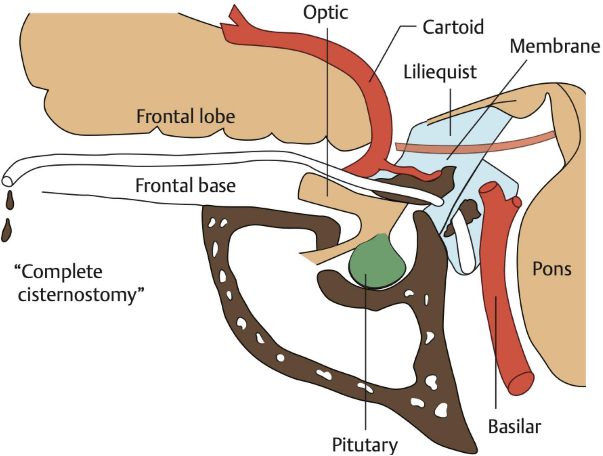
Figure 1. Schematic of basal cistern anatomy demonstrating carotid, chiasmatic, interpeduncular, and prepontine compartments with the Liliequist membrane.
Indications and contraindications
Indications include severe TBI (GCS 3–8) with effaced cisterns, refractory ICP (>20 mmHg despite tiered therapy), and aneurysmal SAH with substantial cisternal clot (5, 6, 10). The technique may be considered when decompressive craniectomy is contemplated but bone preservation is desirable. Contraindications include established brainstem failure, bilateral fixed dilated pupils, significant coagulopathy, and haemodynamic instability.
Operative technique
Stepwise microsurgical cisternostomy demonstrating Sylvian fissure dissection, exposure of the opticocarotid and chiasmatic cisterns, and progressive cerebrospinal fluid (CSF) release leading to brain relaxation mentioned in Video 1.
| Video 1. Stepwise microsurgical cisternostomy demonstrating Sylvian fissure dissection, exposure of the opticocarotid and chiasmatic cisterns, and progressive cerebrospinal fluid (CSF) release leading to brain relaxation. Key anatomical landmarks and arachnoid membranes are highlighted to illustrate safe and effective decompression. https://youtu.be/k9d-QQcuHrU |
Anaesthesia and monitoring
General anaesthesia is administered with attention to maintaining cerebral perfusion pressure. Normocapnia, normothermia, and haemodynamic stability are targeted. An external ventricular drain (EVD) allows ICP monitoring and controlled CSF diversion (3).
Positioning
The patient is positioned supine with 15–20° contralateral head rotation and slight extension. Rigid fixation is used. This configuration promotes gravity-assisted frontal lobe relaxation and reduces the need for fixed retraction.
Craniotomy and skull base preparation
A tailored pterional craniotomy is performed. Generous drilling of the sphenoid wing flattens the skull base, shortens the working corridor, and improves the angle of approach, thereby minimising retraction.
Extradural release
Extradural steps enhance exposure: division of the orbitomeningeal band, temporal dural peeling, and selective anterior clinoidectomy where indicated. These manoeuvres increase surgical freedom and decrease retraction-related risk (11).
Dural opening
A basal dural opening directed towards the sphenoid ridge facilitates early access to the Sylvian fissure and basal cisterns. The dura may be loosely approximated at closure to accommodate postoperative swelling.
Sylvian fissure dissection
Wide Sylvian fissure splitting is undertaken along arachnoid planes under high magnification. The M1 segment of the middle cerebral artery serves as a reliable guide to the carotid cistern. Preservation of Sylvian veins is essential to avoid venous infarction. Wide Sylvian fissure dissection with exposure of the carotid cistern is demonstrated in Figure 2.
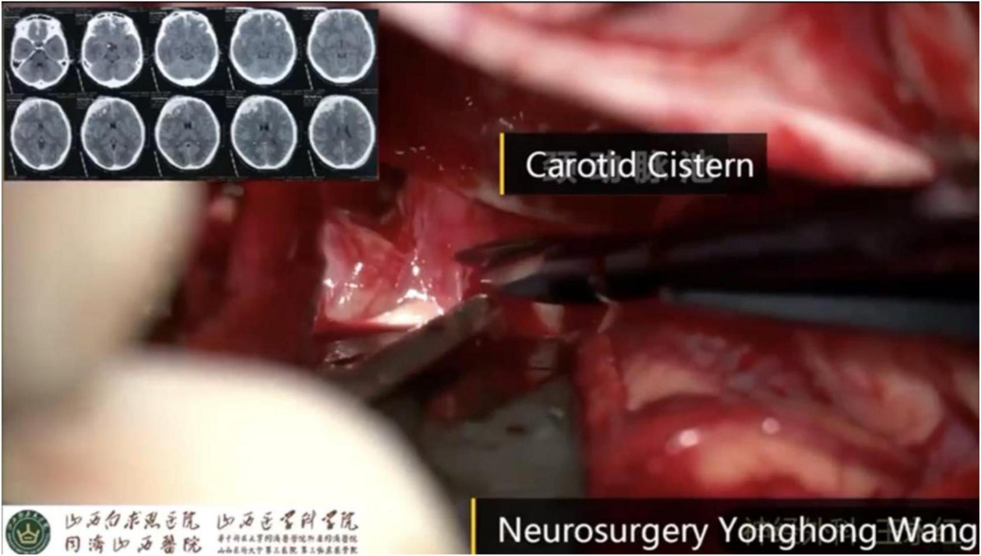
Figure 2. Wide Sylvian fissure dissection showing exposure of the carotid cistern and arachnoid plane development.
Carotid cistern opening
Opening the carotid cistern typically produces the first substantial CSF egress and visible brain relaxation. Key landmarks include the internal carotid artery, optic nerve, and A1 segment. Opening of the carotid cistern results in initial cerebrospinal fluid egress, as shown in Figure 3.
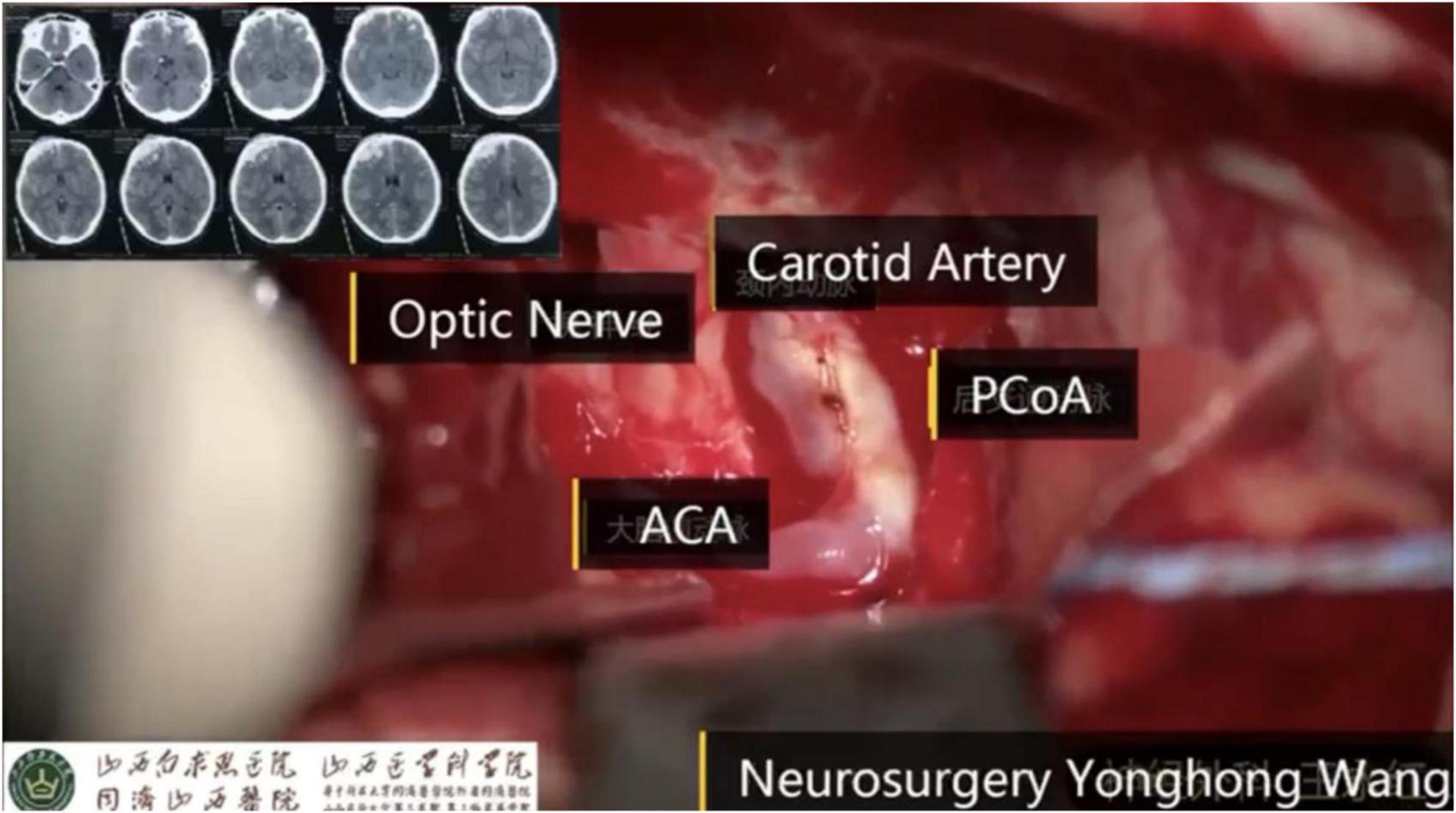
Figure 3. Carotid cistern opening with identification of the internal carotid artery and optic nerve, demonstrating initial CSF egress.
Chiasmatic cistern opening
Medial extension of the dissection opens the chiasmatic cistern, exposing the optic chiasm and anterior communicating artery complex. In SAH, dense adhesions are common and are best managed with sharp arachnoid dissection. Further medial dissection exposes the chiasmatic cistern and optic apparatus (Figure 4).
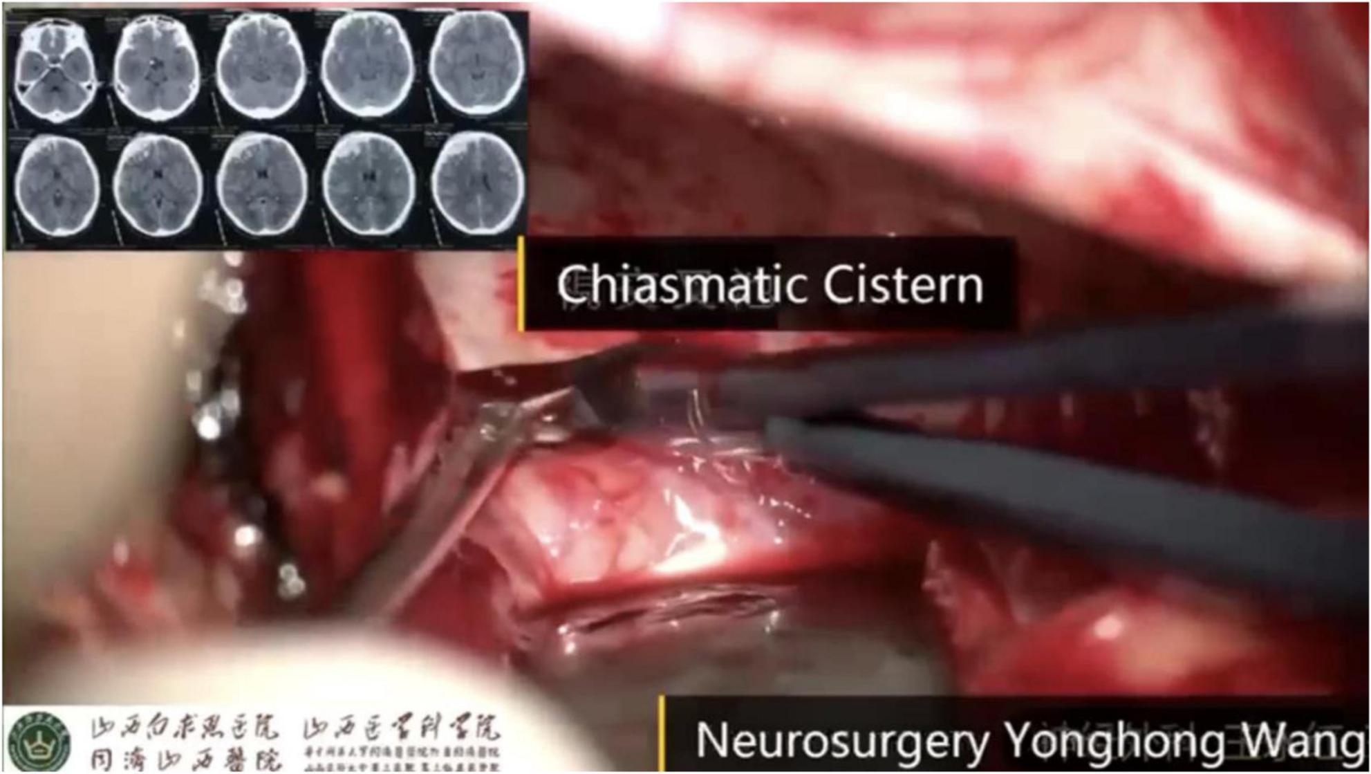
Figure 4. Chiasmatic cistern exposure with optic chiasm and anterior communicating artery complex.
Liliequist membrane fenestration
Fenestration of the Liliequist membrane is pivotal. The oculomotor nerve must be identified prior to incision. Both diencephalic and mesencephalic leaves are opened to establish communication with the prepontine cistern; the basilar artery becomes visible when entry is adequate (7, 9). Fenestration of the Liliequist membrane, before and after opening, is illustrated in Figure 5.
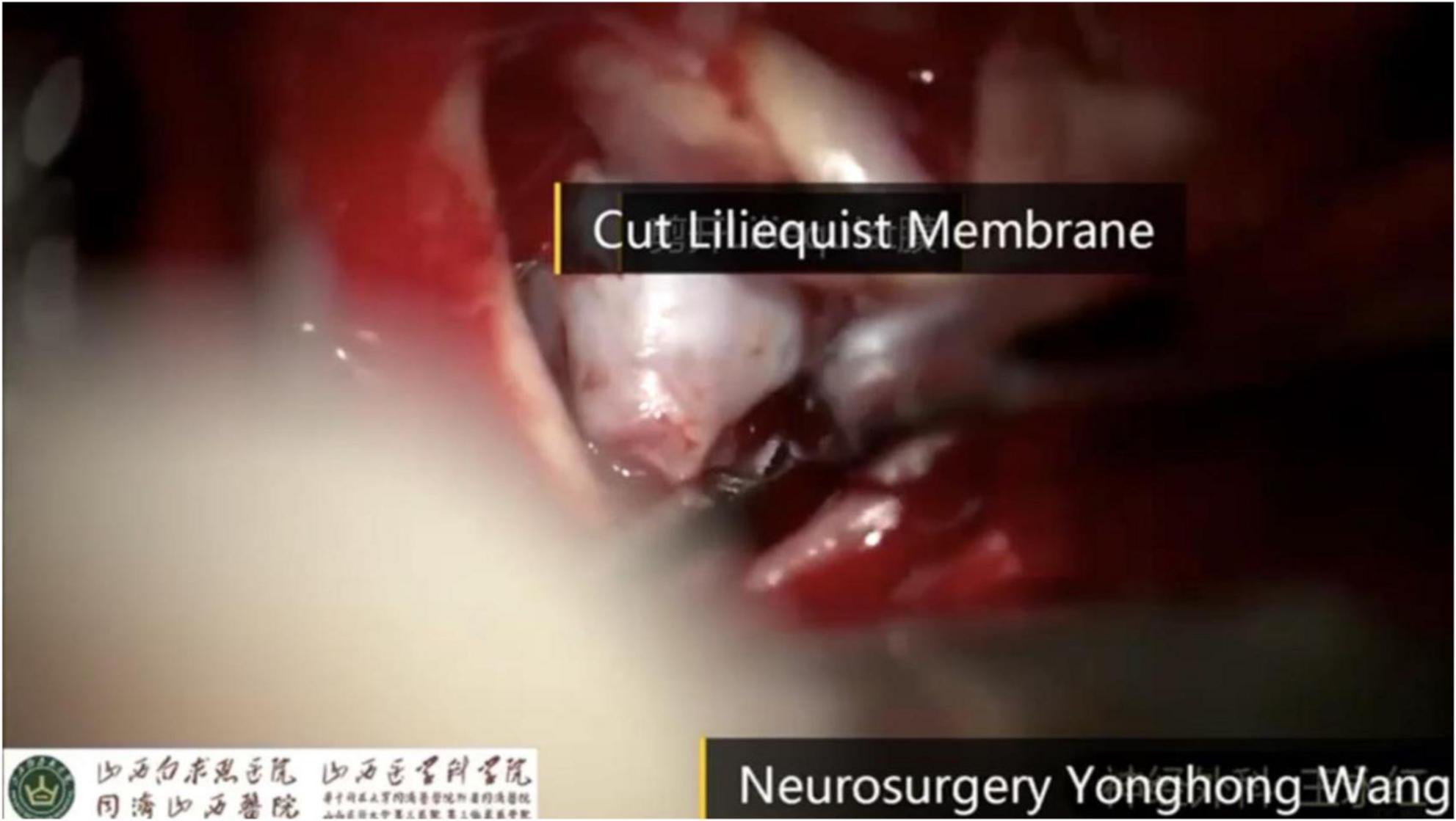
Figure 5. Liliequist membrane before and after fenestration with entry into the prepontine cistern and basilar artery visualisation.
Prepontine cistern and drain placement
After membrane opening, the prepontine cistern is accessed. A soft silastic catheter is placed under direct vision along the ventral brainstem to enable continuous CSF drainage and, when required, gentle irrigation (10). Final placement of the cisternal drain within the prepontine cistern is demonstrated in Figure 6.
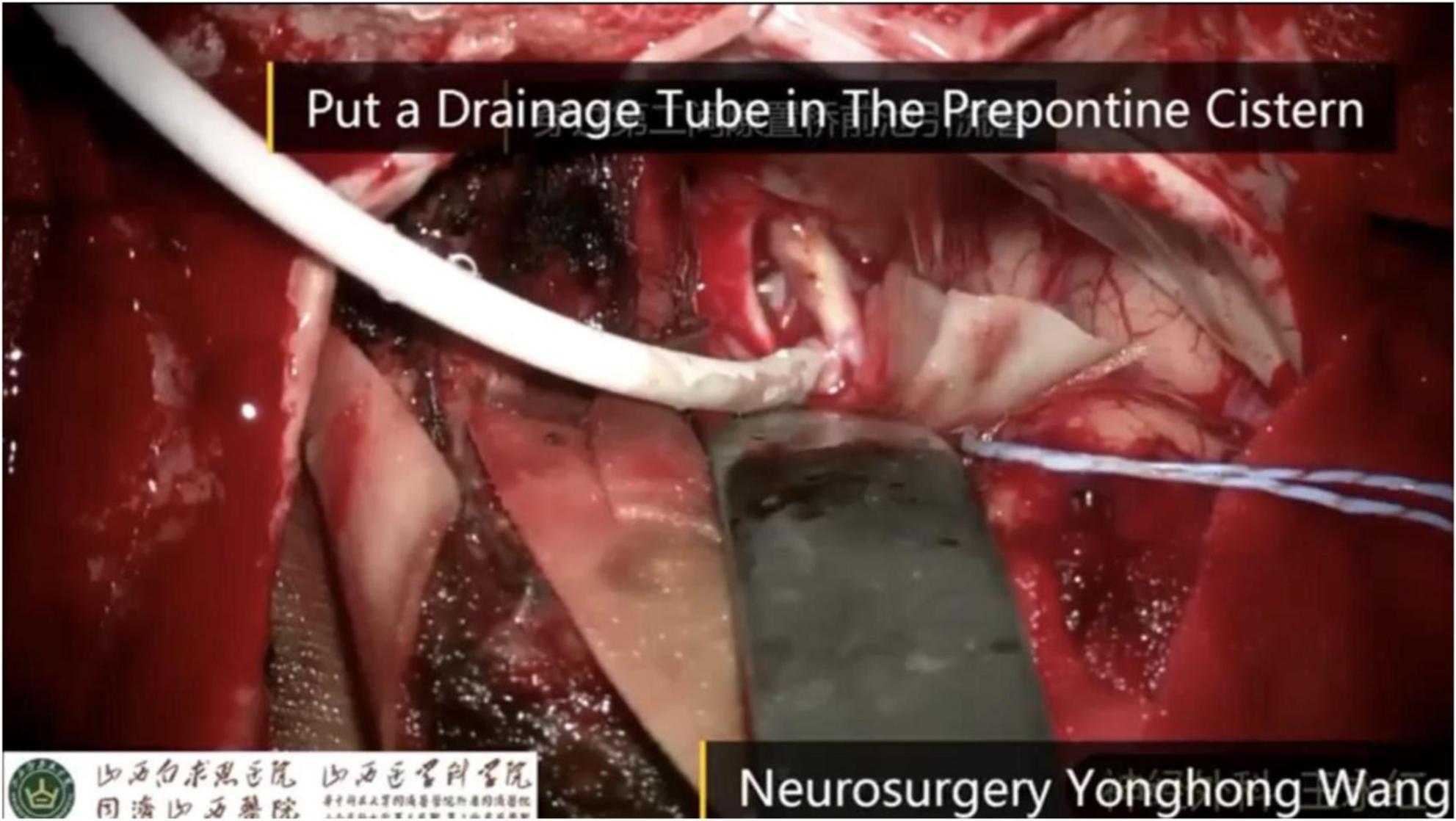
Figure 6. Prepontine cisternal drain in situ along the ventral brainstem.
Closure
Meticulous haemostasis is achieved and the field is irrigated. The cisternal drain is secured. The dura is loosely approximated, and the bone flap is replaced to preserve cranial integrity while allowing expansion.
Surgical nuances and technical pearls
Brain relaxation should be used as the operative endpoint; persistent tension suggests incomplete decompression. All manoeuvres must remain within arachnoid planes, avoiding traction on neural or vascular structures. Perforating vessels around the carotid bifurcation and basilar apex require careful preservation to prevent infarction (12). In SAH, clot removal should be gradual with irrigation and gentle aspiration. Cranial nerve safety is paramount—particularly the oculomotor nerve during membrane fenestration and the abducens nerve in the prepontine region. Proper positioning and maintenance of the cisternal drain are essential for sustained postoperative effect.
Clinical evidence
Prospective and comparative studies report effective ICP reduction, decreased requirement for decompressive craniectomy, and improved functional outcomes following cisternostomy in selected cohorts (1, 2, 5, 6). In SAH, early cisternal clearance may reduce vasospasm-related complications (10, 13). While high-quality multicentre data are still evolving, current evidence supports a physiological benefit when the technique is performed meticulously.
Conclusion
Cisternostomy directly addresses impaired CSF dynamics by restoring communication across basal cisterns. When executed with careful anatomical orientation and microsurgical discipline, it can achieve durable ICP control without removing the skull. Mastery of wide Sylvian fissure opening, sequential cisternal decompression, and complete Liliequist membrane fenestration is essential for consistent results.
Funding
The authors received no financial support for the research, authorship, and/or publication of this article.
Conflict of interest
The authors declare that the research was conducted in the absence of any commercial or financial relationships that could be construed as a potential conflict of interest.
References
1. Jiang Y, Lv H, Huang S et al. Cisternostomy for traumatic brain injury: a systematic review and meta-analysis. Neurosurg Rev. (2022) 45(1):251–62.
2. Singh G, Cherian I, Bhattarai B et al. Cisternostomy for traumatic brain injury: a meta-analysis of clinical outcomes. World Neurosurg. (2021) 149:e390–8.
3. Cherian I, Yi G, Munakomi S. Cisternostomy: replacing decompressive hemicraniectomy? Asian J Neurosurg. (2013) 8(3):132–8.
4. Iliff JJ, Wang M, Liao Y, Plogg BA, Peng W, Gundersen GA et al. A paravascular pathway facilitates CSF flow through the brain parenchyma. Sci Transl Med. (2012) 4(147):147ra111.
5. Cherian I, Bhattarai B, Grasso G et al. Cisternostomy for intracranial pressure management in traumatic brain injury. Surg Neurol Int. (2017) 8:10.
6. Cherian I, Bernardo A, Grasso G. Cisternostomy in traumatic brain injury: pathophysiologic mechanisms and surgical technique. World Neurosurg. (2016) 89:51–7.
7. Matsuno H, Rhoton AL Jr., Peace D. Microsurgical anatomy of the posterior fossa cisterns. Neurosurgery. (1988) 23(1):58–80.
8. Grasso G, Torregrossa F, Cherian I. Cisternostomy for traumatic brain injury: anatomical and physiological considerations. Brain Sci. (2021) 11(7):836.
10. Cherian I, Bhattarai B, Grasso G. Cisternostomy as an adjunct to surgery for ruptured aneurysm. Acta Neurochir (Wien). (2014) 156(8):1523–5.
11. Rosenørn J, Diemer N. The risk of cerebral damage during graded brain retractor pressure in the rat. J Neurosurg. (1985) 63(4):608–11.
13. Macdonald RL, Schweizer TA. Spontaneous subarachnoid haemorrhage. Lancet. (2017) 389(10069):655–66.
© The Author(s). 2026 Open Access This article is distributed under the terms of the Creative Commons Attribution 4.0 International License (https://creativecommons.org/licenses/by/4.0/), which permits unrestricted use, distribution, and reproduction in any medium, provided you give appropriate credit to the original author(s) and the source, provide a link to the Creative Commons license, and indicate if changes were made.