Case presentation
Patient history
A 45-year-old female (Patient X) presented with a 3-month history of left-sided, intermittent clear watery nasal discharge. The rhinorrhea was described as continuous, unilateral, and characteristically increased on forward bending or Valsalva manoeuvre — features highly suggestive of CSF rhinorrhea. There was no antecedent history of head trauma, prior sinonasal surgery, or infectious sinusitis. No neurological deficits were noted on examination. General physical examination was unremarkable, and the patient was normotensive.
Diagnostic investigations
Computed tomography — paranasal sinuses (high-resolution CT)
High-resolution CT of the paranasal sinuses with multiplanar reconstruction was performed to evaluate bony anatomy and identify skull base defects. Key findings included:
• Mild deviation of the nasal septum to the left with a left-sided bony septal spur
• Mild hypertrophy of the right inferior turbinate
• Focal bony defect measuring approximately 2 mm in the left cribriform plate
• Subtle adjacent fluid density in the region of the defect, suggestive of CSF leak
• No evidence of sinonasal mass, polyp, or inflammatory sinus disease
MRI cisternography
MRI cisternography was performed using high-resolution T2-weighted sequences to characterise the defect and confirm active CSF communication. Salient findings included:
• Defect measuring approximately 2 mm in the left cribriform plate at the ethmoid roof, located at the junction of the nasal septum and middle turbinate attachment
• Signal characteristics consistent with CSF tracking into the nasal cavity through the identified defect
• Empty sella, indicative of chronically elevated intracranial pressure — a key finding raising suspicion for IIH as the aetiology of the spontaneous leak (1)
• Mild diffuse cerebral atrophy
• Periventricular white matter hyperintensities consistent with chronic microangiopathic changes
Patient was then planned for Trans Nasal Trans Endoscopic CSF leak repair surgery (2, 3).
Surgical technique
All procedures were performed under general anaesthesia with the patient in the supine position, head elevated 15–20 degrees. Intravenous prophylactic antibiotics (Cefazolin 2 g) were administered 30 minutes prior to incision. Neuronavigation using CT and MRI fusion data was available for intraoperative guidance. The following steps were executed in sequence.
Step 1: Anterior septoplasty and correction of septal deviation
A Killian incision was made at the mucocutaneous junction of the left nasal vestibule. Subperichondrial and subperiosteal dissection was carried out bilaterally along the septal framework. The deviated quadrilateral cartilage was scored and repositioned.
The bovie electrocautery with a fine-tipped bovine electrode was used to facilitate controlled subperichondrial dissection and haemostasis throughout the septoplasty, enabling precise tissue handling with minimal mucosal trauma.
Step 2: Endoscopic identification and delineation of the CSF leak
A 0-degree rigid nasal endoscope (4 mm) was introduced via the left nasal cavity. Under direct endoscopic visualisation, the skull base was systematically inspected.
The meningocele/encephalocele sac prolapsing through the defect was identified and carefully cauterised using bipolar electrocautery at low power to devitalise the herniated tissue without thermal injury to adjacent intracranial structures. Following cauterisation and shrinkage of the herniated tissue, the bony defect was precisely delineated using a Freer elevator and a fine-tipped suction. The defect was confirmed to measure 8 mm x 3 mm, straddling the attachment of the middle turbinate — a transversely oriented defect that extended from the lateral lamella across the cribriform plate.
Step 3: Partial middle turbinectomy
To ensure adequate exposure of the skull base defect and to create a flat, accessible platform for the multilayer repair, the anterior one-third to one-half of the left middle turbinate was excised using through-cutting forceps and Blakesley forceps. Care was taken to preserve the posterior lamella and turbinate attachment to avoid destabilisation of the middle turbinate remnant. The turbinectomy revealed the full extent of the bony defect spanning the cribriform plate and confirmed its dimensions and configuration.
The edges of the bony defect were freshened circumferentially with a diamond burr to remove residual mucosa, creating a raw, vascular surface that would promote adhesion and integration of the grafts.
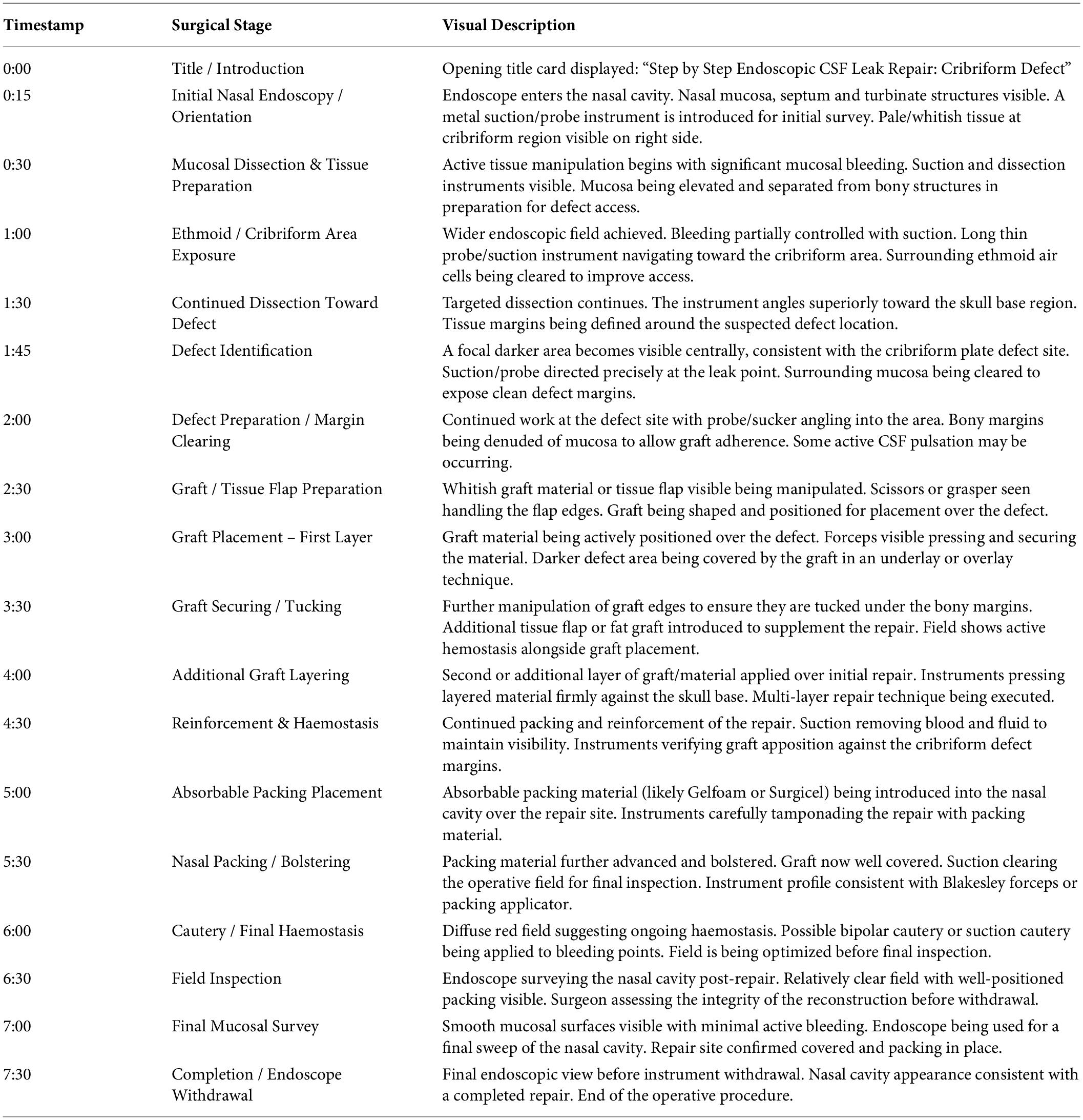
Table 1. Post operative events were uneventful. Post extubation, nasal packing was done and patient was stable. After 2 days, nasal packing was removed and no CSF leak was observed.
Step 4: Multilayer reconstruction — underlay fascia lata
A 3 x 3 cm piece of fascia lata was harvested from the patient’s left thigh through a small longitudinal incision. Adequate graft dimensions exceeding the skull base defect by a minimum of 5 mm on all margins were ensured. The graft was trimmed to the appropriate size and hydrated.
The first layer of fascia lata was introduced into the nasal cavity with alligator forceps and positioned as an intracranial underlay. The graft was carefully manoeuvred under endoscopic visualisation through the bony defect, ensuring it lay flat against the intradural surface. Gentle pressure was applied to confirm stable seating of the underlay component.
Step 5: Cartilage gasket placement
A piece of septal cartilage obtained during the prior septoplasty was carved and shaped to fit snugly within the bony defect. The cartilage gasket was sized to be approximately 0.5 mm smaller than the defect dimensions on each side to permit insertion while achieving a firm press-fit when seated. The cartilage was placed within the bony defect in direct apposition with the underlay fascia lata, effectively sandwiching the dura between the intracranial underlay and the extracranial cartilage. This gasket creates a mechanical seal analogous to a flanged plug and is the hallmark of the gasket-seal technique (4).
Step 6: Overlay fascia lata
A second, larger piece of fascia lata was then placed as an extracranial overlay over the cartilage gasket and the surrounding bony rim of the defect. This overlay layer was positioned to extend well beyond the bony margins in all four directions, providing complete soft tissue coverage. The overlay fascia was inspected to confirm circumferential contact with the freshened bony edges, ensuring a watertight secondary seal.
Step 7: Elevation and inset of the nasoseptal flap
Prior to the repair steps, at the onset of the procedure, the right-sided mucoperichondrial and mucoperiosteal nasoseptal flap had been elevated based on the posterior septal artery — a branch of the sphenopalatine artery. The flap was elevated from the posterior septum from just posterior to the columella anteriorly to the sphenoid face posteriorly, and from the floor of the nasal cavity inferiorly to the level of the olfactory strip superiorly. The posterior pedicle was preserved intact to maintain robust vascular supply (5).
The fully elevated nasoseptal flap was then rotated superiorly and medially to cover the repair site. The flap was draped over the overlay fascia lata and the cartilage gasket, ensuring complete coverage of the defect and its reconstructed margins. The flap edges were apposed to the surrounding mucosal margins and secured using fibrin glue (Tisseel) applied circumferentially to reinforce the seal and promote adhesion (5).
Post operative events were uneventful. Post extubation, nasal packing was done and patient was stable. After 2 days, nasal packing was removed and no CSF leak was observed.
Step-by-Step Transnasal Endoscopic Cerebrospinal Fluid Leak Repair: Cribriform Defect with Multilayer Gasket-Seal Closure and Nasoseptal Flap Reconstruction. [2026]. Video file. Duration: 7 min 41 sec. Resolution: 1920×1080, 24 fps.
| Video 1. Pre-operative high-resolution CT of the paranasal sinuses (coronal and sagittal multiplanar reconstructions) demonstrating a focal bony defect in the left cribriform plate with adjacent fluid density, followed by MRI cisternography confirming active CSF communication and radiological markers of raised intracranial pressure (empty sella). Intraoperative endoscopic footage documents the sequential steps of transnasal endoscopic CSF leak repair: anterior septoplasty via Killian incision with cartilage harvest, endoscopic identification and cauterisation of the herniated meningocele sac, partial middle turbinectomy for defect exposure, intracranial underlay placement of fascia lata, press-fit autologous cartilage gasket insertion, extracranial overlay fascia lata placement, and inset of a posteriorly pedicled nasoseptal flap secured with fibrin glue. Total operative footage duration: approximately 7 minutes 41 seconds. https://youtu.be/Acj_xoiRrWY |
Conflict of interest
The author declares that the research was conducted in the absence of any commercial or financial relationships that could be construed as a potential conflict of interest.
References
1. Woodworth BA, Prince A, Chiu AG, Cohen NA, Schlosser RJ, Bolger WE, et al. Spontaneous CSF leaks: a paradigm for definitive repair and management of intracranial hypertension. Otolaryngol Head Neck Surg. 2008;138(6):715–720. doi: 10.1016/j.otohns.2008.02.010
2. Lobo BC, Baumanis MM, Nelson RF. Surgical repair of spontaneous cerebrospinal fluid (CSF) rhinorrhea: systematic review and meta-analysis. Laryngoscope Investig Otolaryngol. 2017;2(5):215–224. doi: 10.1002/lio2.73
3. Psaltis AJ, Schlosser RJ, Banks CA, Yawn J, Soler ZM. A systematic review of the endoscopic repair of cerebrospinal fluid rhinorrhea. Laryngoscope. 2012;122(3):555–560. doi: 10.1002/lary.22488
4. Leng LZ, Brown S, Anand VK, Schwartz TH. “Gasket-seal” watertight closure in minimal-access endoscopic cranial base surgery. Neurosurgery. 2008;62(5 Suppl 2):ONSE342–343. doi: 10.1227/01.neu.0000316273.18052.72
5. Hadad G, Bassagasteguy L, Carrau RL, Mataza JC, Kassam A, Snyderman CH et al. A novel reconstructive technique after endoscopic expanded endonasal approaches: vascular pedicle nasoseptal flap. Laryngoscope. 2006;116(10):1882–1886. doi: 10.1097/01.mlg.0000234933.37779.e4
© The Author(s). 2026 Open Access This article is distributed under the terms of the Creative Commons Attribution 4.0 International License (https://creativecommons.org/licenses/by/4.0/), which permits unrestricted use, distribution, and reproduction in any medium, provided you give appropriate credit to the original author(s) and the source, provide a link to the Creative Commons license, and indicate if changes were made.